|
|
Expandable Metal Stents for the Treatment of
Cancerous Obstruction of the Gastrointestinal Tract
Todd H. Baron, M.D. Expandable metal stents have been approved by the Food and
Drug Administration for the treatment of gastrointestinal
obstruction due to cancer. Although they have not been approved for
use in benign disease, there are specific clinical indications for
which expandable metal stents may be beneficial. This article reviews
the uses of expandable metal stents for gastrointestinal obstruction
due to cancer.
General Concepts
Gastrointestinal stents are placed by gastroenterologists under
endoscopic guidance with the aid of fluoroscopy or by interventional
radiologists using fluoroscopic guidance alone. Expandable metal stents
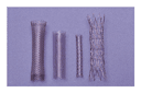
are made of metal alloys and have varying shapes and sizes,
depending on the manufacturer and the organ in which they will be
placed (Figure 1).
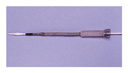
The stents are mounted in a preloaded constrained position on a
delivery catheter (Figure
2). A guide wire is passed through the lumen of the catheter,
and when the wire has been advanced beyond the obstruction, the
stent is passed over it and positioned across the stricture. The
constraining mechanism is released, which causes the length of the
stent to decrease and its diameter to increase (Figure 2). The
radial expansile forces and the degree of shortening differ among
different types of stents.1
Covered metal stents have a membrane to prevent reobstruction due to
ingrowth of tumor through the mesh wall.
|
|
Specimens obtained from animals2 and
from humans at autopsy or surgery3
show that metal stents embed themselves in the tumor and surrounding
tissue with pressure necrosis and are incorporated into the wall of
the organ. Completely covered stents do not become embedded, and as
a result, migration of the stent is possible. Therefore, partially
covered stents are used to prevent tumor ingrowth and permit
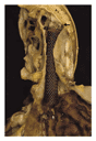
anchoring of the stent (Figure 2). One
of the possible adverse effects is erosion through the gastrointestinal wall
(Figure 3).
Most metal stents appear safe for patients undergoing magnetic
resonance imaging (MRI); specific details of the stent and its
orientation to the magnetic field should be obtained before MRI is
performed.4,5
|
Esophageal Stents
Esophageal carcinoma accounts for most cases of dysphagia due
to cancer, and usually the tumor is unresectable. Dysphagia may
also result from extrinsic compression due to lung cancer or
malignant lymphadenopathy. Among many endoscopic and nonendoscopic treatment
alternatives for palliation of dysphagia due to cancer, expandable
metal stents are one of the main options. They are useful for
patients with poor functional status who cannot tolerate radiation
or chemotherapy, who have advanced metastatic disease, or in whom
previous therapy has failed.6
The small diameter of expandable metal stents before deployment
makes aggressive dilation of the esophagus before or after deployment
unnecessary. Despite the substantially higher cost of expandable metal
stents as compared with traditional rigid plastic esophageal stents,
there are substantial overall cost savings resulting from the
reduction in the number of days of hospitalization due to
complications.7,8,9 One
study showed that the rate of complications associated with stent
insertion was lower overall for expandable metal stents than for
plastic stents, but the rate of subacute complications was higher.10
In the United States, expandable metal stents have replaced plastic
stents for use in the esophagus.11
Dysphagia is relieved in approximately 90 percent of patients who
receive expandable metal stents.7,8,9
The data from comparisons of different options for the palliation
of dysphagia due to cancer are limited. A retrospective study compared
expandable metal stents with a variety of conventional endoscopic
palliative techniques in patients with inoperable esophageal
carcinoma without tracheoesophageal fistula. Patients with expandable
metal stents underwent significantly fewer procedures and spent
fewer days in the hospital.12
In a prospective, randomized, controlled trial of patients with
esophageal carcinoma, patients with expandable metal stents had
significantly more improvement in symptoms and a lower rate of
reintervention than those treated by esophageal laser
recanalization.13
An advantage of expandable metal stents over other endoscopic
palliative methods is that they can be used to treat dysphagia due
to compression caused by cancer,14,15
although the improvement in dysphagia is less than for patients with
esophageal cancer.14
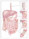
Esophageal expandable metal stents are also used to treat
tracheoesophageal fistulas due to cancer (Figure 4A).16,17
Tracheoesophageal fistulas develop in patients with advanced
esophageal and lung cancer and lead to continuous aspiration of
saliva. Tracheoesophageal fistula is the only condition in which
covered expandable metal stents may increase survival as compared
with other therapies. Although there have been no prospective trials
comparing covered metal stents with other types for the treatment of
tracheoesophageal fistulas, the covered metal stent is now accepted
as the primary treatment option. Closure of the fistula is
successful in 70 to 100 percent of patients.16,17
For persistent fistulas, placement of an airway stent to close the
fistula18
(Figure 4A)
or surgical esophageal bypass for physiologically fit patients19
are additional palliative options.
|
Expandable metal stents are best suited for midesophageal lesions. Tumors
at the gastroesophageal junction are amenable to stent placement
with a high rate of technical and clinical success, but stents at
this location produce an open conduit for free reflux of gastric
contents, with consequent severe regurgitation and aspiration.11
Newer stents prevent reflux and aspiration by means of a one-way
flap valve on the gastric side of the stent.20
Placement of expandable metal stents for very proximal esophageal
lesions is technically difficult because of the proximity of the
stent to the upper esophageal sphincter and the lack of an
uninvolved proximal margin. However, in studies of small numbers of
patients with dysphagia due to esophageal obstruction caused by
cancer high in the cervical esophagus, expandable metal stents were
successfully placed and improved symptoms in most patients.21
Placement of an esophageal expandable metal stent can lead to
severe complications.11
Intraprocedural complications include those associated with
conscious sedation, aspiration, malpositioning of the stent, and
esophageal perforation. Immediate postprocedural complications may
include chest pain, bleeding, and tracheal compression, with
resultant airway compromise and respiratory arrest. Late
complications include distal stent migration, formation of an
esophageal fistula, bleeding, perforation, and stent occlusion. Although
most migrated stents can be retrieved endoscopically or will simply
pass through the gastrointestinal tract, small-bowel obstruction
develops in some patients. Approximately 0.5 to 2 percent of
patients who undergo the procedure die as a direct result of
placement of an expandable metal stent (Figure 3).11
Several studies strongly suggest that the rates of delayed
esophageal complications caused by expandable metal stents are
higher in patients who have previously been treated with radiation,
chemotherapy, or both.6,22,23
These complications are presumably due to stent-induced pressure
necrosis within devitalized esophageal tissue. Unfortunately, patients
who have recurrent or persistent dysphagia or a tracheoesophageal fistula
after chemoradiation therapy often have no alternative to a stent
for palliation of their symptoms. In contrast to other studies, one
prospective study found that patients undergoing chemoradiation
therapy after placement of an expandable metal stent had
significantly longer survival than patients who received only a
stent.24
However, shrinkage of the tumor by chemoradiation therapy could
increase the risk of stent migration.
Patients with esophageal stents must modify their diet to prevent
large boluses of food from becoming impacted within the stent. If
a stent without an antireflux valve crosses the gastroesophageal junction,
strict antireflux precautions and aggressive acid suppression are
needed to prevent gastroesophageal reflux and aspiration. If the
stent lumen becomes occluded by tissue ingrowth, overgrowth, or
hyperplasia, a second stent may be placed through the first stent
with good results.25
Alternatively, endoscopic laser therapy, electrocautery, or
photodynamic therapy may be used to treat stent occlusion.
Biliary Stents
Treatment of obstructive jaundice due to cancer relieves pruritus,
improves appetite, and reduces fat malabsorption.26
Surgical palliation of this condition involves the creation of an
anastomosis between the bile duct and the duodenum or jejunum to
bypass the obstructed biliary tree. Nonsurgical palliation is
achieved by placing stents endoscopically (with the use of
endoscopic retrograde cholangiopancreatography), or with radiologic
guidance (by the percutaneous transhepatic approach) across the cancerous
stricture to restore biliary continuity (Figure 4B).
Insertion of plastic biliary stents provides an effective
alternative to open palliative surgical bypass of the biliary tree
for the management of obstructive jaundice due to cancer.27,28
Unfortunately, bacterial encrustation frequently leads to occlusion
of plastic stents,29
requiring their replacement in terminally ill patients. Plastic
stents of larger diameter take longer to become occluded.30
The maximal stent diameter is limited by the endoscopes that must
accommodate them and the size of the tract made through the liver
when they are placed percutaneously. In comparison with plastic
stents, expandable metal biliary stents have a smaller diameter
before placement and a larger diameter after placement. Their small
diameter before placement makes percutaneous placement less
traumatic and permits them to be completely placed during one
procedure. Their large diameter after placement eliminates occlusion
by bacterial biofilm.
Expandable metal biliary stents are much more costly than plastic
stents. In two randomized, prospective trials comparing endoscopically
placed, uncovered metal biliary stents and plastic stents for the
palliation of jaundice in patients with previously untreated distal
malignant bile-duct obstruction, the metal stents had significantly
longer patency.31,32
Decreases in the rates of endoscopic procedures and
rehospitalization offset the initial high cost of metal stents.
Similar results were found in a large prospective, randomized trial
in which stents were placed by the percutaneous transhepatic route
for palliation of distal cancerous biliary obstruction.33
Expandable metal stents are more cost effective for patients who
survive longer than four to six months, whereas a single procedure
with placement of a less costly plastic stent would suffice in
patients expected to have shorter survival.34,35
Another disadvantage of a metal stent in the biliary tree, as
compared with a plastic stent, is the fact that the device cannot be
removed once it is implanted. Therefore, in patients with
potentially resectable cancer, removable plastic stents should be
used.
Expandable metal biliary stents may become occluded because of
tumor ingrowth or biliary epithelial hyperplasia induced by the
stent. Occlusion from either cause may be treated by the insertion
of a plastic stent or another metal stent through the original
stent.31,35,36
Preliminary studies suggest that occlusion due to tumor ingrowth or
epithelial hyperplasia may be prevented by the use of partially
covered metal biliary stents.37
Overgrowth of tumor beyond the ends of the stent may result in
reobstruction, necessitating placement of another stent.
Prolonged patency has been observed in expandable biliary stents
used to treat low- or distal-bile-duct obstruction, but not in
cases of obstruction by a tumor involving the proximal biliary system
at or above the bifurcation of the right and left hepatic ducts
(Klatskin's tumor). However, one small randomized trial and several
uncontrolled studies suggest that expandable metal stents are more
effective than plastic stents in these locations as well.38
Complications related to expandable metal biliary stents include
entrapment of the stent-delivery system in small or nondilated intrahepatic
ducts, malpositioning of the stent, and trauma to the duodenal wall
opposite the papilla, with resultant bleeding or perforation if an
excessive amount of the distal portion of the stent protrudes into
the lumen. There is no apparent increase in complications in
patients who have previously received chemoradiation therapy or who
receive it concomitantly.39
Although expandable metal stents are generally not removable, there
are case reports of successful endoscopic removal of such biliary
stents for the treatment of late complications.
Gastroduodenal Stents
Successful placement of expandable metal stents for palliation
of cancerous obstruction of the upper gastrointestinal tract has
been reported in several series, with clinical success rates similar
to those of surgical palliative bypass.40,41,42
Approximately 90 percent of patients with gastroduodenal stents
improve clinically.40
Advanced carcinoma of the pancreatic head is the most common cancer
that obstructs the gastric outlet (Figure 4C).
Gastric carcinoma or disease that metastasizes to the duodenum or
jejunum may also cause obstruction.43
Patients with cancerous duodenal obstruction often also have biliary
obstruction that occurs first.43
Given the difficulties in obtaining access to the biliary tree
through the mesh wall of a duodenal stent placed across the papilla,
an expandable metal biliary stent should be placed before the
duodenal stent is placed if there is known or impending biliary
obstruction. Bile flows effectively through the biliary and duodenal
stents as they cross within the duodenum (Figure 4C).
To treat biliary obstruction after placement of a duodenal stent,
a percutaneous transhepatic approach is usually required. Stenting
of both the duodenum and the bile duct is the nonsurgical equivalent
of a traditional double surgical bypass.
Endoscopic placement of gastroduodenal stents under fluoroscopic
guidance is technically easier than their placement by interventional
radiologists using fluoroscopic guidance alone, because the obstruction
can be reached directly and because there is a mechanical advantage
in passing the stent through the endoscope channel. Successful
relief of obstruction in the proximal jejunum can be achieved
endoscopically.43
Patients may resume oral intake almost immediately after
uncomplicated placement of expandable metal stents in the upper
gastrointestinal tract. They should be advised to advance from
liquids to solids as tolerated and to avoid leafy vegetables, which
may result in stent occlusion. Gastroduodenal stents are often
placed in outpatient procedures.
Complications after placement of expandable metal stents in the
upper gastrointestinal tract include perforation, bleeding, stent
migration, stent malpositioning, and occlusion of the stent by tumor
overgrowth or ingrowth or by food impaction. The condition of some
patients with advanced cancer and gastroduodenal obstruction will
not improve after successful stent placement because of
gastrointestinal obstruction due to tumor at other, unidentified
sites, diffuse peritoneal carcinomatosis with bowel encasement, or
functional gastric-outlet obstruction due to neural involvement by
tumor (e.g., of the celiac axis).43
There are no data about the safety of expandable metal stents in the
stomach or small bowel in patients who have already received or
are currently receiving chemoradiation therapy.
Colorectal Stents
Placement of a colorectal stent should be considered for
preoperative decompression and for palliation of cancerous large-bowel
obstruction.40
Up to 30 percent of patients with primary colorectal carcinoma present
with large-bowel obstruction.40
The traditional method of managing complete or subtotal colonic
obstruction due to cancer, particularly left-sided obstruction,
involves the creation of a diverting colostomy. Patients with
complete or subtotal colonic obstruction and a potentially
resectable tumor cannot undergo a one-stage operative resection of
the tumor and immediate reanastomosis, because stool within the
uncleansed proximal colon leads to breakdown of the anastomosis.
Therefore, the initial surgery includes resection of the primary
tumor and colostomy, with reanastomosis at a second operation.
Patients with complete colonic obstruction tend to be acutely ill,
with advanced disease. Because preoperative placement of an
expandable metal colorectal stent permits clinical stabilization
with preoperative decompression and cleansing, a one-stage operation
can then be performed and colostomy avoided.44,45
The stent is removed en bloc at the time of resection of the primary
tumor, after serving as a bridge to surgery. If the patient is a
poor candidate for surgical resection because of underlying illness
or has unresectable or widely metastatic disease discovered by
imaging studies, the stent can remain in place for palliation. A
recent multicenter study of patients with primary colon carcinoma
evaluated the effectiveness of preoperative placement of metal
stents inserted radiologically.45
Successful stent placement, with clinical resolution of large-bowel
obstruction within 96 hours, was achieved in 66 of 71 patients (93
percent). Sixty-five patients underwent elective single-stage
surgery with a primary colonic anastomosis a mean of 8.6 days after
stent placement. A severe complication (intestinal perforation)
occurred in one patient.
In a retrospective study of the management of acute cancerous
colonic obstruction, consecutive patients with colorectal carcinoma who
received expandable metal stents were compared with a similar group
of consecutive patients who underwent traditional surgical treatment
at the same institution.46
When the data for patients who subsequently underwent a curative
resection were analyzed, a cost savings of 28.8 percent was seen in
the group receiving stents, because of decreases in the total number
of days in the hospital, the number of days in the intensive care
unit, and the number of surgical procedures. Despite these promising
results, no completed randomized, prospective studies have compared preoperative
stents with standard surgery in a group of patients with potentially
resectable primary colorectal cancer and obstruction. One such study
is under way in the United States. It remains to be seen whether
long-term results, such as tumor-recurrence rates, are altered by
the use of preoperative placement of colorectal stents.
Candidates for placement of a colorectal stent for palliation
include patients with colorectal carcinoma and obstruction who have
extensive local or metastatic disease or who are poor candidates for
surgical resection, and patients with colonic obstruction secondary
to noncolonic pelvic cancers (e.g., bladder or ovarian carcinoma) or
metastatic cancer (e.g., breast carcinoma) (Figure 4D).
44,47,48
Successful palliation of obstruction with avoidance of colostomy can
be achieved in 85 to 100 percent of patients, with some stents
remaining patent and in place for more than one year.48,49
Randomized trials are needed to confirm these findings. At present,
however, it is difficult to deny such patients the option of
receiving a stent in order to avoid a permanent colostomy. Partially
covered colonic stents have also been used to close cancerous
colovesical and colovaginal fistulas, with an acceptable risk of
stent migration.50
Complications of the placement of colonic stents include
perforation, stent migration, bleeding, stent malpositioning, and
occlusion of the stent by stool. Colonic perforation during
insertion of colonic stents may be devastating, because fecal
material is spilled into the abdominal cavity, resulting in
peritonitis. The peritonitis may be difficult to manage surgically,
because the patient may become even more acutely ill, potentially
worsening the surgical outcome. Stents placed low in the rectum may
produce tenesmus or fecal incontinence.
Stents may be placed endoscopically in the right colon,46
whereas with radiologic guidance alone, stent placement is limited
to the left colon. Patients with widespread advanced cancer may
not have clinical improvement after successful placement of a
colonic stent because of obstruction at other sites or peritoneal carcinomatosis.44
After receiving a palliative colorectal stent, patients should
consume a low-residue diet and use stool softeners or laxatives to
prevent stool impaction and stent occlusion. The effects of previous
or concomitant chemoradiation therapy on rates of local
complications are unknown.
Future Directions
Biodegradable and bioabsorbable expandable stents are being developed
for the treatment of benign disease.51
Such stents may be useful in treating gastrointestinal strictures
that are refractory to dilation, such as peptic esophageal
strictures, anastomotic or radiation-induced strictures, or
strictures related to Crohn's disease. In patients with cancer, the
use of expandable metal stents that emit radiation52
or release chemotherapeutic agents53
may cause tumor regression.
Source Information
From the Department of Medicine, Division of Gastroenterology and
Hepatology, Mayo Foundation, Rochester, Minn.
Address reprint requests to Dr. Baron at 200 First St. SW,
Eisenberg 8A, Rochester, MN 55905, or at [log in to unmask].
References
- Chan
AC, Shin FG, Lam YH, et al. A comparison study on physical properties of
self-expandable esophageal metal stents. Gastrointest Endosc
1999;49:462-465.
[Medline]
- Silvis
SE, Sievert CE Jr, Vennes JA, Abeyta BK, Brennecke LH. Comparison of
covered versus uncovered wire mesh stents in the canine biliary tract.
Gastrointest Endosc 1994;40:17-21.
[Medline]
- Bethge
N, Sommer A, Gross U, von Kleist D, Vakil N. Human tissue responses to
metal stents implanted in vivo for the palliation of malignant stenoses.
Gastrointest Endosc 1996;43:596-602.
[Medline]
- Taal BG,
Muller SH, Boot H, Koops W. Potential risks and artifacts of magnetic
resonance imaging of self-expandable esophageal stents. Gastrointest
Endosc 1997;46:424-429.
[Medline]
- Nitatori
T, Hanaoka H, Hachiya J, Yokoyama K. MRI artifacts of metallic stents
derived from imaging sequencing and the ferromagnetic nature of materials.
Radiat Med 1999;17:329-334.
[Medline]
- Bethge
N, Sommer A, von Kleist D, Vakil N. A prospective trial of self-expanding
metal stents in the palliation of malignant esophageal obstruction after
failure of primary curative therapy. Gastrointest Endosc 1996;44:283-286.
[Medline]
- Knyrim
K, Wagner H-J, Bethge N, Keymling M, Vakil N. A controlled trial of an
expansile metal stent for palliation of esophageal obstruction due to
inoperable cancer. N Engl J Med 1993;329:1302-1307.
[Abstract/Full Text]
- De Palma
GD, di Matteo E, Romano G, Fimmano A, Rondinone G, Catanzano C. Plastic
prosthesis versus expandable metal stents for palliation of inoperable
esophageal thoracic carcinoma: a controlled prospective study.
Gastrointest Endosc 1996;43:478-482.
[Medline]
- Roseveare
CD, Patel P, Simmonds N, Goggin PM, Kimble J, Shepherd HA. Metal stents
improve dysphagia, nutrition and survival in malignant oesophageal
stenosis: a randomized controlled trial comparing modified Gianturco
Z-stents with plastic Atkinson tubes. Eur J Gastroenterol Hepatol
1998;10:653-657.
[Medline]
- Kozarek
RA, Ball TJ, Brandabur JJ, et al. Expandable versus conventional
esophageal prostheses: easier insertion may not preclude subsequent
stent-related problems. Gastrointest Endosc 1996;43:204-208.
[Medline]
- Ramirez
FC, Dennert B, Zierer ST, Sanowski RA. Esophageal self-expandable metallic
stents -- indications, practice, techniques, and complications: results of
a national survey. Gastrointest Endosc 1997;45:360-364.
[Medline]
- Nicholson
DA, Haycox A, Kay CL, Rate A, Attwood S, Bancewicz J. The cost
effectiveness of metal oesophageal stenting in malignant disease compared
with conventional therapy. Clin Radiol 1999;54:212-215.
[Medline]
- Adam A,
Ellul J, Watkinson AF, et al. Palliation of inoperable esophageal
carcinoma: a prospective randomized trial of laser therapy and stent
placement. Radiology 1997;202:344-348.
[Abstract]
- Bethge
N, Sommer A, Vakil N. Palliation of malignant esophageal obstruction due
to intrinsic and extrinsic lesions with expandable metal stents. Am J
Gastroenterol 1998;93:1829-1832.
[Medline]
- Gupta
NK, Boylan CE, Razzaq R, England RE, Mirra L, Martin DF. Self-expanding
oesophageal metal stents for the palliation of dysphagia due to extrinsic
compression. Eur Radiol 1999;9:1893-1897.
[Medline]
- Morgan
RA, Ellul JP, Denton ER, Glynos M, Mason RC, Adam A. Malignant esophageal
fistulas and perforations: management with plastic-covered metallic
endoprostheses. Radiology 1997;204:527-532.
[Medline]
- Raijman
I, Siddique I, Ajani J, Lynch P. Palliation of malignant dysphagia and
fistulae with coated expandable metal stents: experience with 101 patients.
Gastrointest Endosc 1998;48:172-179.
[Medline]
- Freitag
L, Tekolf E, Steveling H, Donovan TJ, Stamatis G. Management of malignant
esophagotracheal fistulas with airway stenting and double stenting. Chest
1996;110:1155-1160.
[Abstract]
- Low DE,
Kozarek RA. Comparison of conventional and wire mesh expandable prostheses
and surgical bypass in patients with malignant esophagorespiratory
fistulas. Ann Thorac Surg 1998;65:919-923.
[Abstract]
- Kocher
M, Dlouhy M, Neoral C, et al. Esophageal stent with antireflux valve for
tumors involving the cardia: work in progress. J Vasc Interv Radiol
1998;9:1007-1010.
[Abstract]
- Bethge
N, Sommer A, Vakil N. A prospective trial of self-expanding metal stents
in the palliation of malignant esophageal strictures near the upper
esophageal sphincter. Gastrointest Endosc 1997;45:300-303.
[Medline]
- Kinsman
KJ, DeGregorio BT, Katon RM, et al. Prior radiation and chemotherapy
increase the risk of life-threatening complications after insertion of
metallic stents for esophagogastric malignancy. Gastrointest Endosc 1996;43:196-203.
[Medline]
- Siersema
PD, Hop WC, Dees J, Tilanus HW, van Blankenstein M. Coated self-expanding
metal stents versus latex prostheses for esophagogastric cancer with
special reference to prior radiation and chemotherapy: a controlled,
prospective study. Gastrointest Endosc 1998;47:113-120.
[Medline]
- Ludwig
D, Dehne A, Burmester E, Wiedemann GJ, Stange EF. Treatment of
unresectable carcinoma of the esophagus or the gastroesophageal junction
by mesh stents with or without radiochemotherapy. Int J Oncol
1998;13:583-588.
[Medline]
- Lagattolla
NR, Rowe PH, Anderson H, Dunk AA. Restenting malignant oesophageal
strictures. Br J Surg 1998;85:261-263.
[Medline]
- Ballinger
AB, McHugh M, Catnach SM, Alstead EM, Clark ML. Symptom relief and quality
of life after stenting for malignant bile duct obstruction. Gut
1994;35:467-470.
[Abstract]
- Smith
AC, Dowsett JF, Russell RC, Hatfield AR, Cotton PB. Randomised trial of
endoscopic stenting versus surgical bypass in malignant low bileduct
obstruction. Lancet 1994;344:1655-1660.
[Medline]
- Speer
AG, Cotton PB, Russell RC, et al. Randomised trial of endoscopic versus
percutaneous stent insertion in malignant obstructive jaundice. Lancet
1987;2:57-62.
[Medline]
- Speer
AG, Cotton PB, Rode J, et al. Biliary stent blockage with bacterial
biofilm: a light and electron microscopy study. Ann Intern Med
1988;108:546-553.
[Medline]
- Speer
AG, Cotton PB, MacRae KD. Endoscopic management of malignant biliary
obstruction: stents of 10 French gauge are preferable to stents of 8
French gauge. Gastrointest Endosc 1988;34:412-417.
[Medline]
- Davids
PH, Groen AK, Rauws EA, Tytgat GN, Huibregtse K. Randomised trial of
self-expanding metal stents versus polyethylene stents for distal
malignant biliary obstruction. Lancet 1992;340:1488-1492.
[Medline]
- Knyrim
K, Wagner HJ, Pausch J, Vakil N. A prospective, randomized, controlled
trial of metal stents for malignant obstruction of the common bile duct.
Endoscopy 1993;25:207-212.
[Medline]
- Lammer
J, Hausegger KA, Fluckiger F, et al. Common bile duct obstruction due to
malignancy: treatment with plastic versus metal stents. Radiology
1996;201:167-172.
[Abstract]
- Yeoh KG,
Zimmerman MJ, Cunningham JT, Cotton PB. Comparative costs of metal versus
plastic biliary stent strategies for malignant obstructive jaundice by
decision analysis. Gastrointest Endosc 1999;49:466-471.
[Medline]
- Prat F,
Chapat O, Ducot B, et al. A randomized trial of endoscopic drainage
methods for inoperable malignant strictures of the common bile duct.
Gastrointest Endosc 1998;47:1-7.
[Medline]
- Tham TC,
Carr-Locke DL, Vandervoort J, et al. Management of occluded biliary
Wallstents. Gut 1998;42:703-707.
[Abstract/Full Text]
- Shim CS,
Lee YH, Cho YD, et al. Preliminary results of a new covered biliary metal
stent for malignant biliary obstruction. Endoscopy 1998;30:345-350.
[Medline]
- Wagner
HJ, Knyrim K, Vakil N, Klose KJ. Plastic endoprostheses versus metal
stents in the palliative treatment of malignant hilar biliary obstruction:
a prospective and randomized trial. Endoscopy 1993;25:213-218.
[Medline]
- Eschelman
DJ, Shapiro MJ, Bonn J, et al. Malignant biliary duct obstruction:
long-term experience with Gianturco stents and combined-modality radiation
therapy. Radiology 1996;200:717-724.
[Abstract]
- Mauro
MA, Koehler RE, Baron TH. Advances in gastrointestinal intervention: the
treatment of gastroduodenal and colorectal obstructions with metallic
stents. Radiology 2000;215:659-669.
[Abstract/Full Text]
- Feretis
C, Benakis P, Dimopoulos C, Manouras A, Tsimbloulis B, Apostolidis N.
Duodenal obstruction caused by pancreatic head carcinoma: palliation with
self-expandable endoprostheses. Gastrointest Endosc 1997;46:161-165.
[Medline]
- Jung GS,
Song HY, Kang SG, et al. Malignant gastroduodenal obstructions: treatment
by means of a covered expandable metallic stent -- initial experience.
Radiology 2000;216:758-763.
[Abstract/Full Text]
- Yates MR
III, Morgan DE, Baron TH. Palliation of malignant gastric and small
intestinal strictures with self-expandable metal stents. Endoscopy
1998;30:266-272.
[Medline]
- Baron
TH, Dean PA, Yates MR III, Canon C, Koehler RE. Expandable metal stents
for the treatment of colonic obstruction: techniques and outcomes.
Gastrointest Endosc 1998;47:277-286.
[Medline]
- Mainar
A, De Gregorio Ariza MA, Tejero E, et al. Acute colorectal obstruction:
treatment with self-expandable metallic stents before scheduled surgery --
results of a multicenter study. Radiology 1999;210:65-69.
[Abstract/Full Text]
- Binkert
CA, Ledermann H, Jost R, Saurenmann P, Decurtins M, Zollikofer CL. Acute
colonic obstruction: clinical aspects and cost-effectiveness of
preoperative and palliative treatment with self-expanding metallic stents
-- a preliminary report. Radiology 1998;206:199-204.
[Abstract]
- de
Gregorio MA, Mainar A, Tejero E, et al. Acute colorectal obstruction:
stent placement for palliative treatment -- results of a multicenter
study. Radiology 1998;209:117-120.
[Abstract]
- Fernandez
Lobato R, Pinto I, Paul L, et al. Self-expanding prostheses as a
palliative method in treating advanced colorectal cancer. Int Surg
1999;84:159-162.
[Medline]
- Camunez
F, Echenagusia A, Simo G, Turegano F, Vazquez J, Barreiro-Meiro I.
Malignant colorectal obstruction treated by means of self-expanding
metallic stents: effectiveness before surgery and in palliation. Radiology
2000;216:492-497.
[Abstract/Full Text]
- Repici
A, Reggio D, De Angelis C, et al. Covered metal stents for management of
inoperable malignant colorectal strictures. Gastrointest Endosc
2000;52:735-740.
[Medline]
- Tamai H,
Igaki K, Kyo E, et al. Initial and 6-month results of biodegradable
poly-l-lactic acid coronary stents in humans. Circulation 2000;102:399-404.
[Abstract/Full Text]
- Zamora
PO, Osaki S, Som P, et al. Radiolabeling brachytherapy sources with Re-188
through chelating microfilms: stents. J Biomed Mater Res 2000;53:244-251.
[Medline]
- Herdeg
C, Oberhoff M, Karsch KR. Antiproliferative stent coatings: Taxol and
related compounds. Semin Interv Cardiol 1998;3:197-199.
[Medline]
Edward E.
Rylander, M.D.
Diplomat American
Board of Family Practice.
Diplomat American
Board of Palliative Medicine.