Dr. Wendy Leu had the first correct answer.
Discussion: Alkaptonuria
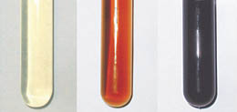
A, Appearance of the urine at the time of the
initial void; B, at 24 hours postvoid; C, at 48 hours postvoid.
Alkaptonuria is a rare disorder of tyrosine
metabolism inherited as an autosomal recessive trait, with an incidence of 1 in
200 000.1 A deficiency of
homogentisic acid oxidase, the cause of this disorder, leads to the
accumulation of large amounts of homogentisic acid in the body with subsequent
excretion of the acid in the urine. The gene responsible for alkaptonuria has
been mapped to the long arm of chromosome 3, 3q2.2
CLINICAL FEATURES
The only clinical sign of this disorder in the pediatric age group is the
darkening of urine to an almost black color on standing. The change in
coloration, the result of oxidation and polymerization of homogentisic acid, is
enhanced in an alkaline urine. An acid urine may not become darkened after
standing many hours.
The major clinical features of alkaptonuria are
not evident until mid adulthood and are the result of deposition of a
blue-black pigment, derived from oxidation of homogentisic acid, in cartilage
and connective tissue. The pigment results in degeneration of cartilage,
particularly that of the spine and large joints (hips and knees). The
arthritis, known as ochronotic arthritis
because of the color of the pigment in cartilage, has clinical characteristics
of rheumatoid arthritis, but radiologic findings are typical of osteoarthritis.2 Stiffness and
discomfort of the back are usually the initial symptoms of the arthritis.
The first evidence of pigment deposition in
cartilage and connective tissue is noted in adults in the third and fourth
decades of life. A faint slate-gray coloration may be perceived through the skin
overlaying the cartilage of the nose and ears.3 Discoloration of the
sclera of the eyes may also be noted. Patients with homogentisic acid oxidase
deficiency have a higher incidence of heart disease, particularly calcification
of the mitral and aortic valves and myocardial infarctions.3
DIAGNOSIS
A presumptive diagnosis of alkaptonuria may be made by demonstrating the change
in color of standing urine over time. Confirmation is made by measuring the
excretion of homogentisic acid in the urine. The addition of oxidizing agents
such as silver nitrate, ferric chloride, or Benedict reagent to the urine will
enhance the color change to brown-black in less than 48 hours.1 Since homogentisic
acid is a strong reducing agent, it will cause a positive reaction with Fehling
or Benedict reagent, whereas it will not result in a positive reaction with
glucose oxidase. Phenol poisoning and malignant melanoma may result in the
passage of dark-colored urine, which is present at the time of voiding rather
than developing on standing.
TREATMENT
There is no specific treatment for alkaptonuria. Theoretically, the deposition
of pigment could be prevented by dietary restriction of phenylalanine and
tyrosine to minimal daily requirements. Large doses of ascorbic acid could
possibly impede oxidation and polymerization of homogentisic acid.
Edward E.
Rylander, M.D.
Diplomat American
Board of Family Practice.
Diplomat American
Board of Palliative Medicine.