The New England Journal of Medicine
Original Article
Volume 345:1368-1377
November 8, 2001
Number 19
Early Goal-Directed Therapy in the Treatment of Severe Sepsis and Septic
Shock
Emanuel Rivers, M.D., M.P.H., Bryant Nguyen, M.D., Suzanne Havstad, M.A.,
Julie Ressler, B.S., Alexandria Muzzin, B.S., Bernhard Knoblich, M.D.,
Edward Peterson, Ph.D., Michael Tomlanovich, M.D., for the Early
Goal-Directed Therapy Collaborative Group
ABSTRACT
Background Goal-directed therapy has been used for severe sepsis and septic
shock in the intensive care unit. This approach involves adjustments of
cardiac preload, afterload, and contractility to balance oxygen delivery
with oxygen demand. The purpose of this study was to evaluate the efficacy
of early goal-directed therapy before admission to the intensive care unit.
Methods We randomly assigned patients who arrived at an urban emergency
department with severe sepsis or septic shock to receive either six hours of
early goal-directed therapy or standard therapy (as a control) before
admission to the intensive care unit. Clinicians who subsequently assumed
the care of the patients were blinded to the treatment assignment.
In-hospital mortality (the primary efficacy outcome), end points with
respect to resuscitation, and Acute Physiology and Chronic Health Evaluation
(APACHE II) scores were obtained serially for 72 hours and compared between
the study groups.
Results Of the 263 enrolled patients, 130 were randomly assigned to early
goal-directed therapy and 133 to standard therapy; there were no significant
differences between the groups with respect to base-line characteristics.
In-hospital mortality was 30.5 percent in the group assigned to early
goal-directed therapy, as compared with 46.5 percent in the group assigned
to standard therapy (P=0.009). During the interval from 7 to 72 hours, the
patients assigned to early goal-directed therapy had a significantly higher
mean (±SD) central venous oxygen saturation (70.4±10.7 percent vs. 65.3±11.4
percent), a lower lactate concentration (3.0±4.4 vs. 3.9±4.4 mmol per
liter), a lower base deficit (2.0±6.6 vs. 5.1±6.7 mmol per liter), and a
higher pH (7.40±0.12 vs. 7.36±0.12) than the patients assigned to standard
therapy (P<=0.02 for all comparisons). During the same period, mean APACHE
II scores were significantly lower, indicating less severe organ
dysfunction, in the patients assigned to early goal-directed therapy than in
those assigned to standard therapy (13.0±6.3 vs. 15.9±6.4, P<0.001).
Conclusions Early goal-directed therapy provides significant benefits with
respect to outcome in patients with severe sepsis and septic shock.
_____
The systemic inflammatory response syndrome can be self-limited or can
progress to severe sepsis and septic shock. 1
<http://content.nejm.org/cgi/content/full/345/19/#R1> Along this continuum,
circulatory abnormalities (intravascular volume depletion, peripheral
vasodilatation, myocardial depression, and increased metabolism) lead to an
imbalance between systemic oxygen delivery and oxygen demand, resulting in
global tissue hypoxia or shock. 2
<http://content.nejm.org/cgi/content/full/345/19/#R2> An indicator of
serious illness, global tissue hypoxia is a key development preceding
multiorgan failure and death. 2
<http://content.nejm.org/cgi/content/full/345/19/#R2> The transition to
serious illness occurs during the critical "golden hours," when definitive
recognition and treatment provide maximal benefit in terms of outcome. These
golden hours may elapse in the emergency department, 3
<http://content.nejm.org/cgi/content/full/345/19/#R3> hospital ward, 4
<http://content.nejm.org/cgi/content/full/345/19/#R4> or the intensive care
unit. 5 <http://content.nejm.org/cgi/content/full/345/19/#R5>
Early hemodynamic assessment on the basis of physical findings, vital signs,
central venous pressure, 6
<http://content.nejm.org/cgi/content/full/345/19/#R6> and urinary output 7
<http://content.nejm.org/cgi/content/full/345/19/#R7> fails to detect
persistent global tissue hypoxia. A more definitive resuscitation strategy
involves goal-oriented manipulation of cardiac preload, afterload, and
contractility to achieve a balance between systemic oxygen delivery and
oxygen demand. 2 <http://content.nejm.org/cgi/content/full/345/19/#R2> End
points used to confirm the achievement of such a balance (hereafter called
resuscitation end points) include normalized values for mixed venous oxygen
saturation, arterial lactate concentration, base deficit, and pH. 8
<http://content.nejm.org/cgi/content/full/345/19/#R8> Mixed venous oxygen
saturation has been shown to be a surrogate for the cardiac index as a
target for hemodynamic therapy. 9
<http://content.nejm.org/cgi/content/full/345/19/#R9> In cases in which the
insertion of a pulmonary-artery catheter is impractical, venous oxygen
saturation can be measured in the central circulation. 10
<http://content.nejm.org/cgi/content/full/345/19/#R10>
Whereas the incidence of septic shock has steadily increased during the past
several decades, the associated mortality rates have remained constant or
have decreased only slightly. 11
<http://content.nejm.org/cgi/content/full/345/19/#R11> Studies of
interventions such as immunotherapy, 12
<http://content.nejm.org/cgi/content/full/345/19/#R12> hemodynamic
optimization, 9 <http://content.nejm.org/cgi/content/full/345/19/#R9> , 13
<http://content.nejm.org/cgi/content/full/345/19/#R13> or pulmonary-artery
catheterization 14 <http://content.nejm.org/cgi/content/full/345/19/#R14>
enrolled patients up to 72 hours after admission to the intensive care unit.
The negative results of studies of the use of hemodynamic variables as end
points ("hemodynamic optimization"), in particular, prompted suggestions
that future studies involve patients with similar causes of disease 13
<http://content.nejm.org/cgi/content/full/345/19/#R13> or with global
tissue hypoxia (as reflected by elevated lactate concentrations) 15
<http://content.nejm.org/cgi/content/full/345/19/#R15> and that they
examine interventions begun at an earlier stage of disease. 16
<http://content.nejm.org/cgi/content/full/345/19/#R16> , 17
<http://content.nejm.org/cgi/content/full/345/19/#R17>
We examined whether early goal-directed therapy before admission to the
intensive care unit effectively reduces the incidence of multiorgan
dysfunction, mortality, and the use of health care resources among patients
with severe sepsis or septic shock.
Methods
Approval of Study Design
This prospective, randomized study was approved by the institutional review
board for human research and was conducted under the auspices of an
independent safety, efficacy, and data monitoring committee.
Eligibility
Eligible adult patients who presented to the emergency department of an
850-bed academic tertiary care hospital with severe sepsis, septic shock, or
the sepsis syndrome from March 1997 through March 2000 were assessed for
possible enrollment according to the inclusion 18
<http://content.nejm.org/cgi/content/full/345/19/#R18> , 19
<http://content.nejm.org/cgi/content/full/345/19/#R19> and exclusion
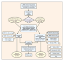
criteria ( Figure 1 <http://content.nejm.org/cgi/content/full/345/19/#F1> ).
The criteria for inclusion were fulfillment of two of four criteria for the
systemic inflammatory response syndrome and a systolic blood pressure no
higher than 90 mm Hg (after a crystalloid-fluid challenge of 20 to 30 ml per
kilogram of body weight over a 30-minute period) or a blood lactate
concentration of 4 mmol per liter or more. The criteria for exclusion from
the study were an age of less than 18 years, pregnancy, or the presence of
an acute cerebral vascular event, acute coronary syndrome, acute pulmonary
edema, status asthmaticus, cardiac dysrhythmias (as a primary diagnosis),
contraindication to central venous catheterization, active gastrointestinal
hemorrhage, seizure, drug overdose, burn injury, trauma, a requirement for
immediate surgery, uncured cancer (during chemotherapy), immunosuppression
(because of organ transplantation or systemic disease), do-not-resuscitate
status, or advanced directives restricting implementation of the protocol.
<http://content.nejm.org/cgi/content/full/345/19/1368/F1>
View larger version (43K):
[in this window] <http://content.nejm.org/cgi/content/full/345/19/1368/F1>
[in a new window]
<http://content.nejm.org/cgi/content-nw/full/345/19/1368/F1>
Figure 1. Overview of Patient Enrollment and Hemodynamic Support.
SIRS denotes systemic inflammatory response syndrome, CVP central venous
pressure, MAP mean arterial pressure, ScvO2 central venous oxygen
saturation, SaO2 arterial oxygen saturation, and VO2 systemic oxygen
consumption. The criteria for a diagnosis of SIRS were temperature greater
than or equal to 38°C or less than 36°C, heart rate greater than 90 beats
per minute, respiratory rate greater than 20 breaths per minute or partial
pressure of arterial carbon dioxide less than 32 mm Hg, and white-cell count
greater than 12,000 per cubic millimeter or less than 4000 per cubic
millimeter or the presence of more than 10 percent immature band forms.
The clinicians who assessed the patients at this stage were unaware of the
patients' treatment assignments. After written informed consent was obtained
(in compliance with the Helsinki Declaration 20
<http://content.nejm.org/cgi/content/full/345/19/#R20> ), the patients were
randomly assigned either to early goal-directed therapy or to standard
(control) therapy in computer-generated blocks of two to eight. The
study-group assignments were placed in sealed, opaque, randomly assorted
envelopes, which were opened by a hospital staff member who was not one of
the study investigators.
Treatment
The patients were treated in a nine-bed unit in the emergency department by
an emergency physician, two residents, and three nurses. 3
<http://content.nejm.org/cgi/content/full/345/19/#R3> The study was
conducted during the routine treatment of other patients in the emergency
department. After arterial and central venous catheterization, patients in
the standard-therapy group were treated at the clinicians' discretion
according to a protocol for hemodynamic support 21
<http://content.nejm.org/cgi/content/full/345/19/#R21> ( Figure 1
<http://content.nejm.org/cgi/content/full/345/19/#F1> ), with critical-care
consultation, and were admitted for inpatient care as soon as possible.
Blood, urine, and other relevant specimens for culture were obtained in the
emergency department before the administration of antibiotics. Antibiotics
were given at the discretion of the treating clinicians. Antimicrobial
therapy was deemed adequate if the in vitro sensitivities of the identified
microorganisms matched the particular antibiotic ordered in the emergency
department. 22 <http://content.nejm.org/cgi/content/full/345/19/#R22>
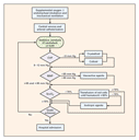
The patients assigned to early goal-directed therapy received a central
venous catheter capable of measuring central venous oxygen saturation
(Edwards Lifesciences, Irvine, Calif.); it was connected to a computerized
spectrophotometer for continuous monitoring. Patients were treated in the
emergency department according to a protocol for early goal-directed therapy
( Figure 2 <http://content.nejm.org/cgi/content/full/345/19/#F2> ) for at
least six hours and were transferred to the first available inpatient beds.
Monitoring of central venous oxygen saturation was then discontinued.
Critical-care clinicians (intensivists, fellows, and residents providing
24-hour in-house coverage) assumed the care of all the patients; these
physicians were unaware of the patients' study-group assignments. The study
investigators did not influence patient care in the intensive care unit.
<http://content.nejm.org/cgi/content/full/345/19/1368/F2>
View larger version (31K):
[in this window] <http://content.nejm.org/cgi/content/full/345/19/1368/F2>
[in a new window]
<http://content.nejm.org/cgi/content-nw/full/345/19/1368/F2>
Figure 2. Protocol for Early Goal-Directed Therapy.
CVP denotes central venous pressure, MAP mean arterial pressure, and ScvO2
central venous oxygen saturation.
The protocol was as follows. A 500-ml bolus of crystalloid was given every
30 minutes to achieve a central venous pressure of 8 to 12 mm Hg. If the
mean arterial pressure was less than 65 mm Hg, vasopressors were given to
maintain a mean arterial pressure of at least 65 mm Hg. If the mean arterial
pressure was greater than 90 mm Hg, vasodilators were given until it was 90
mm Hg or below. If the central venous oxygen saturation was less than 70
percent, red cells were transfused to achieve a hematocrit of at least 30
percent. After the central venous pressure, mean arterial pressure, and
hematocrit were thus optimized, if the central venous oxygen saturation was
less than 70 percent, dobutamine administration was started at a dose of 2.5
µg per kilogram of body weight per minute, a dose that was increased by 2.5
µg per kilogram per minute every 30 minutes until the central venous oxygen
saturation was 70 percent or higher or until a maximal dose of 20 µg per
kilogram per minute was given. Dobutamine was decreased in dose or
discontinued if the mean arterial pressure was less than 65 mm Hg or if the
heart rate was above 120 beats per minute. To decrease oxygen consumption,
patients in whom hemodynamic optimization could not be achieved received
mechanical ventilation and sedatives.
Outcome Measures
The patients' temperature, heart rate, urine output, blood pressure, and
central venous pressure were measured continuously for the first 6 hours of
treatment and assessed every 12 hours for 72 hours. Arterial and venous
blood gas values (including central venous oxygen saturation measured by in
vitro co-oximetry; Nova Biomedical, Waltham, Mass.), lactate concentrations,
and coagulation-related variables and clinical variables required for
determination of the Acute Physiology and Chronic Health Evaluation (APACHE
II) score (on a scale from 0 to 71, with higher scores indicating more
severe organ dysfunction), 23
<http://content.nejm.org/cgi/content/full/345/19/#R23> the Simplified Acute
Physiology Score II (SAPS II, on a scale from 0 to 174, with higher scores
indicating more severe organ dysfunction), 24
<http://content.nejm.org/cgi/content/full/345/19/#R24> and the Multiple
Organ Dysfunction Score (MODS, on a scale from 0 to 24, with higher scores
indicating more severe organ dysfunction) 25
<http://content.nejm.org/cgi/content/full/345/19/#R25> were obtained at
base line (0 hours) and at 3, 6, 12, 24, 36, 48, 60, and 72 hours. 2
<http://content.nejm.org/cgi/content/full/345/19/#R2> , 26
<http://content.nejm.org/cgi/content/full/345/19/#R26> The results of
laboratory tests required only for purposes of the study were made known
only to the study investigators. Patients were followed for 60 days or until
death. The consumption of health care resources (indicated by the duration
of vasopressor therapy and mechanical ventilation and the length of the
hospital stay) was also examined.
Statistical Analysis
In-hospital mortality was the primary efficacy end point. Secondary end
points were the resuscitation end points, organ-dysfunction scores,
coagulation-related variables, administered treatments, and the consumption
of health care resources. Assuming a rate of refusal or exclusion of 10
percent, a two-sided type I error rate of 5 percent, and a power of 80
percent, we calculated that a sample size of 260 patients was required to
permit the detection of a 15 percent reduction in in-hospital mortality.
KaplanMeier estimates of mortality, along with risk ratios and 95 percent
confidence intervals, were used to describe the relative risk of death.
Differences between the two groups at base line were tested with the use of
Student's t-test, the chi-square test, or Wilcoxon's rank-sum test.
Incremental analyses of the area under the curve were performed to quantify
differences during the interval from base line to six hours after the start
of treatment. For the data at six hours, analysis of covariance was used
with the base-line values as the covariates. Mixed models were used to
assess the effect of treatment on prespecified secondary variables during
the interval from 7 to 72 hours after the start of treatment. 27
<http://content.nejm.org/cgi/content/full/345/19/#R27> An independent,
12-member external safety, efficacy, and data monitoring committee reviewed
interim analyses of the data after one third and two thirds of the patients
had been enrolled and at both times recommended that the trial be continued.
To adjust for the two interim analyses, the alpha spending function of
DeMets and Lan 28 <http://content.nejm.org/cgi/content/full/345/19/#R28>
was used to determine that a P value of 0.04 or less would be considered to
indicate statistical significance.
Results
Base-Line Characteristics
We evaluated 288 patients; 8.7 percent were excluded or did not consent to
participate. The 263 patients enrolled were randomly assigned to undergo
either standard therapy or early goal-directed therapy; 236 patients
completed the initial six-hour study period. All 263 were included in the
intention-to-treat analyses. The patients assigned to standard therapy
stayed a significantly shorter time in the emergency department than those
assigned to early goal-directed therapy (mean [±SD], 6.3±3.2 vs. 8.0±2.1
hours; P<0.001). There was no significant difference between the groups in
any of the base-line characteristics, including the adequacy and duration of
antibiotic therapy ( Table 1
<http://content.nejm.org/cgi/content/full/345/19/#T1> ). Vital signs,
resuscitation end points, organ-dysfunction scores, and coagulation-related
variables were also similar in the two study groups at base line ( Table 2
<http://content.nejm.org/cgi/content/full/345/19/#T2> ).
View this table:
[in this window] <http://content.nejm.org/cgi/content/full/345/19/1368/T1>
[in a new window]
<http://content.nejm.org/cgi/content-nw/full/345/19/1368/T1>
Table 1. Base-Line Characteristics of the Patients.
View this table:
[in this window] <http://content.nejm.org/cgi/content/full/345/19/1368/T2>
[in a new window]
<http://content.nejm.org/cgi/content-nw/full/345/19/1368/T2>
Table 2. Vital Signs, Resuscitation End Points, Organ-Dysfunction Scores,
and Coagulation Variables.
Twenty-seven patients did not complete the initial six-hour study period (14
assigned to standard therapy and 13 assigned to early goal-directed
therapy), for the following reasons: discontinuation of aggressive medical
treatment (in 5 patients in each group), discontinuation of aggressive
surgical treatment (in 2 patients in each group), a need for immediate
surgery (in 4 patients assigned to standard therapy and in 3 assigned to
early goal-directed therapy), a need for interventional urologic,
cardiologic, or angiographic procedures (in 2 patients in each group), and
refusal to continue participation (in 1 patient in each group) (P=0.99 for
all comparisons). There were no significant differences between the patients
who completed the initial six-hour study period and those who did not in any
of the base-line characteristics or base-line vital signs, resuscitation end
points, organ-dysfunction scores, or coagulation-related variables (data not
shown).
Vital Signs and Resuscitation End Points
During the initial six hours after the start of therapy, there was no
significant difference between the two study groups in the mean heart rate
(P=0.25) or central venous pressure (P=0.22) ( Table 2
<http://content.nejm.org/cgi/content/full/345/19/#T2> ). During this period,
the mean arterial pressure was significantly lower in the group assigned to
standard therapy than in the group assigned to early goal-directed therapy
(P<0.001), but in both groups the goal of 65 mm Hg or higher was met by all
the patients. The goal of 70 percent or higher for central venous oxygen
saturation was met by 60.2 percent of the patients in the standard-therapy
group, as compared with 94.9 percent of those in the early-therapy group
(P<0.001). The combined hemodynamic goals for central venous pressure, mean
arterial pressure, and urine output (with adjustment for patients with
end-stage renal failure) were achieved in 86.1 percent of the
standard-therapy group, as compared with 99.2 percent of the early-therapy
group (P<0.001). During this period, the patients assigned to standard
therapy had a significantly lower central venous oxygen saturation (P<0.001)
and a greater base deficit (P=0.006) than those assigned to early
goal-directed therapy; the two groups had similar lactate concentrations
(P=0.62) and similar pH values (P=0.26).
During the period from 7 to 72 hours after the start of treatment, the
patients assigned to standard therapy had a significantly higher heart rate
(P=0.04) and a significantly lower mean arterial pressure (P<0.001) than the
patients assigned to early goal-directed therapy; the two groups had a
similar central venous pressure (P=0.68). During this period, those assigned
to standard therapy also had a significantly lower central venous oxygen
saturation than those assigned to early goal-directed therapy (P<0.001), as
well as a higher lactate concentration (P=0.02), a greater base deficit
(P<0.001), and a lower pH (P<0.001).
Organ Dysfunction and Coagulation Variables
During the period from 7 to 72 hours, the APACHE II score, SAPS II, and MODS
were significantly higher in the patients assigned to standard therapy than
in the patients assigned to early goal-directed therapy (P<0.001 for all
comparisons) ( Table 2
<http://content.nejm.org/cgi/content/full/345/19/#T2> ). During this period,
the prothrombin time was significantly greater in the patients assigned to
standard therapy than in those assigned to early goal-directed therapy
(P=0.001), as was the concentration of fibrin-split products (P<0.001) and
the concentration of D-dimer (P=0.006). The two groups had a similar
partial-thromboplastin time (P=0.06), fibrinogen concentration (P=0.21), and
platelet count (P=0.51) ( Table 2
<http://content.nejm.org/cgi/content/full/345/19/#T2> ).
Mortality
In-hospital mortality rates were significantly higher in the
standard-therapy group than in the early-therapy group (P=0.009), as was the
mortality at 28 days (P=0.01) and 60 days (P=0.03) ( Table 3
<http://content.nejm.org/cgi/content/full/345/19/#T3> ). The difference
between the groups in mortality at 60 days primarily reflected the
difference in in-hospital mortality. Similar results were obtained after
data from the 27 patients who did not complete the initial six-hour study
period were excluded from the analysis (data not shown). The rate of
in-hospital death due to sudden cardiovascular collapse was significantly
higher in the standard-therapy group than in the early-therapy group
(P=0.02); the rate of death due to multiorgan failure was similar in the two
groups (P=0.27).
View this table:
[in this window] <http://content.nejm.org/cgi/content/full/345/19/1368/T3>
[in a new window]
<http://content.nejm.org/cgi/content-nw/full/345/19/1368/T3>
Table 3. KaplanMeier Estimates of Mortality and Causes of In-Hospital
Death.
Administered Treatments
During the initial six hours, the patients assigned to early goal-directed
therapy received significantly more fluid than those assigned to standard
therapy (P<0.001) and more frequently received red-cell transfusion
(P<0.001) and inotropic support (P<0.001), whereas similar proportions of
patients in the two groups required vasopressors (P=0.62) and mechanical
ventilation (P=0.90) ( Table 4
<http://content.nejm.org/cgi/content/full/345/19/#T4> ). During the period
from 7 to 72 hours, however, the patients assigned to standard therapy
received significantly more fluid than those assigned to early goal-directed
therapy (P=0.01) and more often received red-cell transfusion (P<0.001) and
vasopressors (P=0.03) and underwent mechanical ventilation (P<0.001) and
pulmonary-artery catheterization (P=0.04); the rate of use of inotropic
agents was similar in the two groups (P=0.14) ( Table 4
<http://content.nejm.org/cgi/content/full/345/19/#T4> ). During the overall
period from base line to 72 hours after the start of treatment, there was no
significant difference between the two groups in the total volume of fluid
administered (P=0.73) or the rate of use of inotropic agents (P=0.15),
although a greater proportion of the patients assigned to standard therapy
than of those assigned to early goal-directed therapy received vasopressors
(P=0.02) and mechanical ventilation (P=0.02) and underwent pulmonary-artery
catheterization (P=0.01), and a smaller proportion required red-cell
transfusion (P<0.001). Though similar between the groups at base line
(P=0.91), the mean hematocrit during this 72-hour period was significantly
lower in the standard-therapy group than in the early-therapy group
(P<0.001). Despite the transfusion of red cells, it was significantly lower
than the value obtained at base line in each group (P<0.001 for both
comparisons) ( Table 2
<http://content.nejm.org/cgi/content/full/345/19/#T2> ).
View this table:
[in this window] <http://content.nejm.org/cgi/content/full/345/19/1368/T4>
[in a new window]
<http://content.nejm.org/cgi/content-nw/full/345/19/1368/T4>
Table 4. Treatments Administered.
Consumption of Health Care Resources
There were no significant differences between the two groups in the mean
duration of vasopressor therapy (2.4±4.2 vs. 1.9±3.1 days, P=0.49), the mean
duration of mechanical ventilation (9.0±13.1 vs. 9.0±11.4 days, P=0.38), or
the mean length of stay in the hospital (13.0±13.7 vs. 13.2±13.8 days,
P=0.54). However, of the patients who survived to hospital discharge, those
assigned to standard therapy had stayed a significantly longer time in the
hospital than those assigned to early goal-directed therapy (18.4±15.0 vs.
14.6±14.5 days, P=0.04).
Discussion
Severe sepsis and septic shock are common and are associated with
substantial mortality and substantial consumption of health care resources.
There are an estimated 751,000 cases (3.0 cases per 1000 population) of
sepsis or septic shock in the United States each year, and they are
responsible for as many deaths each year as acute myocardial infarction
(215,000, or 9.3 percent of all deaths). 29
<http://content.nejm.org/cgi/content/full/345/19/#R29> In elderly persons,
the incidence of sepsis or septic shock and the related mortality rates are
substantially higher than those in younger persons. The projected growth of
the elderly population in the United States will contribute to an increase
in incidence of 1.5 percent per year, yielding an estimated 934,000 and
1,110,000 cases by the years 2010 and 2020, respectively. 29
<http://content.nejm.org/cgi/content/full/345/19/#R29> The present annual
cost of this disease is estimated to be $16.7 billion. 29
<http://content.nejm.org/cgi/content/full/345/19/#R29>
The transition from the systemic inflammatory response syndrome to severe
sepsis and septic shock involves a myriad of pathogenic changes, including
circulatory abnormalities that result in global tissue hypoxia. 1
<http://content.nejm.org/cgi/content/full/345/19/#R1> , 2
<http://content.nejm.org/cgi/content/full/345/19/#R2> These pathogenic
changes have been the therapeutic target of previous outcome studies. 12
<http://content.nejm.org/cgi/content/full/345/19/#R12> Although this
transition occurs over time, both out of the hospital and in the hospital,
in outcome studies interventions have usually been initiated after admission
to the intensive care unit. 12
<http://content.nejm.org/cgi/content/full/345/19/#R12> In studies of
goal-directed hemodynamic optimization, in particular, there was no benefit
in terms of outcome with respect to normal and supranormal hemodynamic end
points, as well as those guided by mixed venous oxygen saturation. 9
<http://content.nejm.org/cgi/content/full/345/19/#R9> , 13
<http://content.nejm.org/cgi/content/full/345/19/#R13> In contrast, even
though we enrolled patients with lower central venous oxygen saturation and
lower central venous pressure than those studied by Gattinoni et al. 9
<http://content.nejm.org/cgi/content/full/345/19/#R9> and with a higher
lactate concentration than those studied by Hayes et al., 13
<http://content.nejm.org/cgi/content/full/345/19/#R13> we found significant
benefits with respect to outcome when goal-directed therapy was applied at
an earlier stage of disease. In patients with septic shock, for example,
Hayes et al. observed a higher in-hospital mortality rate with aggressive
hemodynamic optimization in the intensive care unit (71 percent) than with
control therapy (52 percent), whereas we observed a lower mortality rate in
patients with septic shock assigned to early goal-directed therapy (42.3
percent) than in those assigned to standard therapy (56.8 percent).
The benefits of early goal-directed therapy in terms of outcome are
multifactorial. The incidence of death due to sudden cardiovascular collapse
in the standard-therapy group was approximately double that in the group
assigned to early goal-directed therapy, suggesting that an abrupt
transition to severe disease is an important cause of early death. The early
identification of patients with insidious illness (global tissue hypoxia
accompanied by stable vital signs) makes possible the early implementation
of goal-directed therapy. If sudden cardiovascular collapse can be
prevented, the subsequent need for vasopressors, mechanical ventilation, and
pulmonary-artery catheterization (and their associated risks) diminishes. In
addition to being a stimulus of the systemic inflammatory response syndrome,
global tissue hypoxia independently contributes to endothelial activation
and disruption of the homeostatic balance among coagulation, vascular
permeability, and vascular tone. 30
<http://content.nejm.org/cgi/content/full/345/19/#R30> These are key
mechanisms leading to microcirculatory failure, refractory tissue hypoxia,
and organ dysfunction. 2
<http://content.nejm.org/cgi/content/full/345/19/#R2> , 30
<http://content.nejm.org/cgi/content/full/345/19/#R30> When early therapy
is not comprehensive, the progression to severe disease may be well under
way at the time of admission to the intensive care unit. 16
<http://content.nejm.org/cgi/content/full/345/19/#R16> Aggressive
hemodynamic optimization and other therapy 12
<http://content.nejm.org/cgi/content/full/345/19/#R12> undertaken
thereafter may be incompletely effective or even deleterious. 13
<http://content.nejm.org/cgi/content/full/345/19/#R13>
The value of measurements of venous oxygen saturation at the right atrium or
superior vena cava (central venous oxygen saturation) instead of at the
pulmonary artery (mixed venous oxygen saturation) has been debated, 31
<http://content.nejm.org/cgi/content/full/345/19/#R31> in particular, when
saturation values are above 65 percent. In patients in the intensive care
unit who have hyperdynamic septic shock, the mixed venous oxygen saturation
is rarely below 65 percent. 32
<http://content.nejm.org/cgi/content/full/345/19/#R32> In contrast, our
patients were examined during the phase of resuscitation in which the
delivery of supplemental oxygen is required (characterized by a decreased
mixed venous oxygen saturation and an increased lactate concentration), when
the central venous oxygen saturation generally exceeds the mixed venous
oxygen saturation. 33 <http://content.nejm.org/cgi/content/full/345/19/#R33>
, 34 <http://content.nejm.org/cgi/content/full/345/19/#R34> The initial
central venous oxygen saturation was less than 50 percent in both study
groups. The mixed venous oxygen saturation is estimated to be 5 to 13
percent lower in the pulmonary artery 33
<http://content.nejm.org/cgi/content/full/345/19/#R33> and 15 percent lower
in the splanchnic bed. 35
<http://content.nejm.org/cgi/content/full/345/19/#R35> Though not
numerically equivalent, these ranges of values are pathologically equivalent
and are associated with high mortality. 32
<http://content.nejm.org/cgi/content/full/345/19/#R32> , 36
<http://content.nejm.org/cgi/content/full/345/19/#R36> Among all the
patients in the current study in whom the goals with respect to central
venous pressure, mean arterial pressure, and urine output during the first
six hours were met, 39.8 percent of those assigned to standard therapy were
still in this oxygen-dependent phase of resuscitation at six hours, as
compared with 5.1 percent of those assigned to early goal-directed therapy.
The combined 56.5 percent in-hospital mortality of this 39.8 percent of
patients, who were at high risk for hemodynamic compromise, is consistent
with the results of previous studies in the intensive care unit. 32
<http://content.nejm.org/cgi/content/full/345/19/#R32> , 36
<http://content.nejm.org/cgi/content/full/345/19/#R36>
In an open, randomized, partially blinded trial, there are unavoidable
interactions during the initial period of the study. As the study
progressed, the patients in the standard-therapy group may have received
some form of goal-directed therapy, reducing the treatment effect. This
reduction may have been offset by the slight but inherent bias resulting
from the direct influence of the investigators on the care of the patients
in the treatment group. The potential period of bias was 9.9±19.5 percent of
the overall hospital stay in the standard-therapy group and 7.2±12.0 percent
of that in the group assigned to early goal-directed therapy (P=0.20). This
interval was minimal in comparison with those in previous studies 9
<http://content.nejm.org/cgi/content/full/345/19/#R9> , 13
<http://content.nejm.org/cgi/content/full/345/19/#R13> because the
clinicians who assumed responsibility for the remainder of hospitalization
were completely blinded to the randomization order.
We conclude that goal-directed therapy provided at the earliest stages of
severe sepsis and septic shock, though accounting for only a brief period in
comparison with the overall hospital stay, has significant short-term and
long-term benefits. These benefits arise from the early identification of
patients at high risk for cardiovascular collapse and from early therapeutic
intervention to restore a balance between oxygen delivery and oxygen demand.
In the future, investigators conducting outcome trials in patients with
sepsis should consider the quality and timing of the resuscitation before
enrollment as an important outcome variable.
Supported by the Henry Ford Health Systems Fund for Research, a Weatherby
Healthcare Resuscitation Fellowship, Edwards Lifesciences (which provided
oximetry equipment and catheters), and Nova Biomedical (which provided
equipment for laboratory assays).
We are indebted to the nurses, residents, senior staff attending physicians,
pharmacists, patient advocates, technicians, and billing and administrative
personnel of the Department of Emergency Medicine; to the nurses and
technicians of the medical and surgical intensive care units; and to the
staff members of the Department of Respiratory Therapy, Department of
Pathology, Department of Medical Records, and Department of Admitting and
Discharge for their patience and their cooperation in making this study
possible.
* The members of the Early Goal-Directed Therapy Collaborative Group are
listed in the Appendix.
<http://content.nejm.org/cgi/content/full/345/19/#RFN1>
Source Information
From the Departments of Emergency Medicine (E.R., B.N., J.R., A.M., B.K.,
M.T.), Surgery (E.R.), Internal Medicine (B.N.), and Biostatistics and
Epidemiology (S.H., E.P.), Henry Ford Health Systems, Case Western Reserve
University, Detroit.
Address reprint requests to Dr. Rivers at the Department of Emergency
Medicine, Henry Ford Hospital, 2799 West Grand Blvd., Detroit, MI 48202, or
at [log in to unmask] <mailto:[log in to unmask]> .
References
1. Rangel-Frausto MS, Pittet D, Costigan M, Hwang T, Davis CS, Wenzel RP.
The natural history of the systemic inflammatory response syndrome (SIRS): a
prospective study. JAMA 1995;273:117-123. [Medline]
<http://content.nejm.org/cgi/external_ref?access_num=7799491&link_type=MED>
2. Beal AL, Cerra FB. Multiple organ failure syndrome in the 1990s: systemic
inflammatory response and organ dysfunction. JAMA 1994;271:226-233.
[Medline]
<http://content.nejm.org/cgi/external_ref?access_num=8080494&link_type=MED>
3. Nguyen HB, Rivers EP, Havstad S, et al. Critical care in the emergency
department: a physiologic assessment and outcome evaluation. Acad Emerg Med
2000;7:1354-1361. [Abstract/Full Text]
<http://content.nejm.org/cgi/ijlink?linkType=ABST&journalCode=aemj&resid=7/1
2/1354>
4. Lundberg JS, Perl TM, Wiblin T, et al. Septic shock: an analysis of
outcomes for patients with onset on hospital wards versus intensive care
units. Crit Care Med 1998;26:1020-1024. [Medline]
<http://content.nejm.org/cgi/external_ref?access_num=9635649&link_type=MED>
5. Lefrant JY, Muller L, Bruelle P, et al. Insertion time of the pulmonary
artery catheter in critically ill patients. Crit Care Med 2000;28:355-359.
[Medline]
<http://content.nejm.org/cgi/external_ref?access_num=10708166&link_type=MED>
6. Rady MY, Rivers EP, Nowak RM. Resuscitation of the critically ill in the
ED: responses of blood pressure, heart rate, shock index, central venous
oxygen saturation, and lactate. Am J Emerg Med 1996;14:218-225. [Medline]
<http://content.nejm.org/cgi/external_ref?access_num=8924150&link_type=MED>
7. Cortez A, Zito J, Lucas CE, Gerrick SJ. Mechanism of inappropriate
polyuria in septic patients. Arch Surg 1977;112:471-476. [Medline]
<http://content.nejm.org/cgi/external_ref?access_num=849154&link_type=MED>
8. Elliott DC. An evaluation of the end points of resuscitation. J Am Coll
Surg 1998;187:536-547. [Medline]
<http://content.nejm.org/cgi/external_ref?access_num=9809573&link_type=MED>
9. Gattinoni L, Brazzi L, Pelosi P, et al. A trial of goal-oriented
hemodynamic therapy in critically ill patients. N Engl J Med
1995;333:1025-1032. [Abstract/Full Text]
<http://content.nejm.org/cgi/ijlink?linkType=ABST&journalCode=nejm&resid=333
/16/1025>
10. Reinhart K, Rudolph T, Bredle DL, Hannemann L, Cain SM. Comparison of
central-venous to mixed-venous oxygen saturation during changes in oxygen
supply/demand. Chest 1989;95:1216-1221. [Abstract]
<http://content.nejm.org/cgi/ijlink?linkType=ABST&journalCode=chest&resid=95
/6/1216>
11. Friedman G, Silva E, Vincent JL. Has the mortality of septic shock
changed with time. Crit Care Med 1998;26:2078-86.
12. Opal SM, Cross AS. Clinical trials for severe sepsis: past failures, and
future hopes. Infect Dis Clin North Am 1999;13:285-297. [Medline]
<http://content.nejm.org/cgi/external_ref?access_num=10340167&link_type=MED>
13. Hayes MA, Timmins AC, Yau EHS, Palazzo M, Hinds CJ, Watson D. Elevation
of systemic oxygen delivery in the treatment of critically ill patients. N
Engl J Med 1994;330:1717-1722. [Abstract/Full Text]
<http://content.nejm.org/cgi/ijlink?linkType=ABST&journalCode=nejm&resid=330
/24/1717>
14. Connors AFJ, Speroff T, Dawson NV, et al. The effectiveness of right
heart catheterization in the initial care of critically ill patients. JAMA
1996;276:889-897. [Medline]
<http://content.nejm.org/cgi/external_ref?access_num=8782638&link_type=MED>
15. Haupt MT. Goal-oriented hemodynamic therapy. N Engl J Med
1996;334:799-799.
16. Hinds C, Watson D. Manipulating hemodynamics and oxygen transport in
critically ill patients. N Engl J Med 1995;333:1074-1075. [Full Text]
<http://content.nejm.org/cgi/ijlink?linkType=FULL&journalCode=nejm&resid=333
/16/1074>
17. Shoemaker WC. Goal-oriented hemodynamic therapy. N Engl J Med
1996;334:799-800.
18. American College of Chest Physicians/Society of Critical Care Medicine
Consensus Conference: definitions for sepsis and organ failure and
guidelines for the use of innovative therapies in sepsis. Crit Care Med
1992;20:864-874. [Medline]
<http://content.nejm.org/cgi/external_ref?access_num=1597042&link_type=MED>
19. Sands KE, Bates DW, Lanken PN, et al. Epidemiology of sepsis syndrome in
8 academic medical centers. JAMA 1997;278:234-240. [Medline]
<http://content.nejm.org/cgi/external_ref?access_num=9218672&link_type=MED>
20. World Medical Association Declaration of Helsinki: ethical principles
for medical research involving human subjects. JAMA 2000;284:3043-3045.
[Medline]
<http://content.nejm.org/cgi/external_ref?access_num=11122593&link_type=MED>
21. Task Force of the American College of Critical Care Medicine, Society of
Critical Care Medicine. Practice parameters for hemodynamic support of
sepsis in adult patients in sepsis. Crit Care Med 1999;27:639-660. [Medline]
<http://content.nejm.org/cgi/external_ref?access_num=10199548&link_type=MED>
22. Kollef MH, Sherman G, Ward S, Fraser VJ. Inadequate antimicrobial
treatment of infections: a risk factor for hospital mortality among
critically ill patients. Chest 1999;115:462-474. [Abstract/Full Text]
<http://content.nejm.org/cgi/ijlink?linkType=ABST&journalCode=chest&resid=11
5/2/462>
23. Knaus WA, Draper EA, Wagner DP, Zimmerman JE. APACHE II: a severity of
disease classification system. Crit Care Med 1985;13:818-829. [Medline]
<http://content.nejm.org/cgi/external_ref?access_num=3928249&link_type=MED>
24. Le Gall JR, Lemeshow S, Saulnier F. A new Simplified Acute Physiology
Score (SAPS II) based on a European/North American multicenter study. JAMA
1993;270:2957-2963. [Erratum, JAMA 1994;271:1321.] [Medline]
<http://content.nejm.org/cgi/external_ref?access_num=8254858&link_type=MED>
25. Marshall JC, Cook DJ, Christou NV, Bernard GR, Sprung CL, Sibbald WJ.
Multiple Organ Dysfunction Score: a reliable descriptor of a complex
clinical outcome. Crit Care Med 1995;23:1638-1652. [Medline]
<http://content.nejm.org/cgi/external_ref?access_num=7587228&link_type=MED>
26. Pittet D, Thievent B, Wenzel RP, Li N, Gurman G, Suter PM. Importance of
pre-existing co-morbidities for prognosis of septicemia in critically ill
patients. Intensive Care Med 1993;19:265-272. [Medline]
<http://content.nejm.org/cgi/external_ref?access_num=8408935&link_type=MED>
27. Rutter CM, Elashoff RM. Analysis of longitudinal data: random
coefficient regression modelling. Stat Med 1994;13:1211-1231. [Medline]
<http://content.nejm.org/cgi/external_ref?access_num=7973203&link_type=MED>
28. DeMets DL, Lan KK. Interim analysis: the alpha spending function
approach. Stat Med 1994;13:1341-1356. [Medline]
<http://content.nejm.org/cgi/external_ref?access_num=7973215&link_type=MED>
29. Angus DC, Linde-Zwirble WT, Lidicker J, Clermont G, Carcillo J, Pinsky
MR. Epidemiology of severe sepsis in the United States: analysis of
incidence, outcome, and associated costs of care. Crit Care Med
2001;29:1303-1310. [Medline]
<http://content.nejm.org/cgi/external_ref?access_num=11445675&link_type=MED>
30. Karimova A, Pinsky DJ. The endothelial response to oxygen deprivation:
biology and clinical implications. Intensive Care Med 2001;27:19-31.
[Medline]
<http://content.nejm.org/cgi/external_ref?access_num=11280633&link_type=MED>
31. Edwards JD, Mayall RM. Importance of the sampling site for measurement
of mixed venous oxygen saturation in shock. Crit Care Med 1998;26:1356-1360.
[Medline]
<http://content.nejm.org/cgi/external_ref?access_num=9710094&link_type=MED>
32. Krafft P, Steltzer H, Hiesmayr M, Klimscha W, Hammerle AF. Mixed venous
oxygen saturation in critically ill septic shock patients: the role of
defined events. Chest 1993;103:900-906. [Abstract]
<http://content.nejm.org/cgi/ijlink?linkType=ABST&journalCode=chest&resid=10
3/3/900>
33. Lee J, Wright F, Barber R, Stanley L. Central venous oxygen saturation
in shock: a study in man. Anesthesiology 1972;36:472-478. [Medline]
<http://content.nejm.org/cgi/external_ref?access_num=4553795&link_type=MED>
34. Scheinman MM, Brown MA, Rapaport E. Critical assessment of use of
central venous oxygen saturation as a mirror of mixed venous oxygen in
severely ill cardiac patients. Circulation 1969;40:165-172. [Medline]
<http://content.nejm.org/cgi/external_ref?access_num=5796787&link_type=MED>
35. Dahn MS, Lange MP, Jacobs LA. Central mixed and splanchnic venous oxygen
saturation monitoring. Intensive Care Med 1988;14:373-378. [Medline]
<http://content.nejm.org/cgi/external_ref?access_num=3403769&link_type=MED>
36. Heiselman D, Jones J, Cannon L. Continuous monitoring of mixed venous
oxygen saturation in septic shock. J Clin Monit 1986;2:237-245. [Medline]
<http://content.nejm.org/cgi/external_ref?access_num=3097269&link_type=MED>
Appendix
The following persons participated in the study: External Safety, Efficacy,
and Data Monitoring Committee: A. Connors (Charlottesville, Va.), S. Conrad
(Shreveport, La.), L. Dunbar (New Orleans), S. Fagan (Atlanta), M. Haupt
(Portland, Oreg.), R. Ivatury (Richmond, Va.), G. Martin (Detroit), D.
Milzman (Washington, D.C.), E. Panacek (Palo Alto, Calif.), M. Rady
(Scottsdale, Ariz.), M. Rudis (Los Angeles), and S. Stern (Ann Arbor,
Mich.); the Early-Goal-Directed-Therapy Collaborative Group: B. Derechyk, W.
Rittinger, G. Hayes, K. Ward, M. Mullen, V. Karriem, J. Urrunaga, M.
Gryzbowski, A. Tuttle, W. Chung, P. Uppal, R. Nowak, D. Powell, T. Tyson, T.
Wadley, G. Galletta, K. Rader, A. Goldberg, D. Amponsah, D. Morris, K.
Kumasi-Rivers, B. Thompson, D. Ander, C. Lewandowski, J. Kahler, K.
Kralovich, H. Horst, S. Harpatoolian, A. Latimer, M. Schubert, M. Fallone,
B. Fasbinder, L. Defoe, J. Hanlon, A. Okunsanya, B. Sheridan, Q. Rivers, H.
Johnson, B. Sessa-Boji, K. Gunnerson, D. Fritz, K. Rivers, S. Moore, D.
Huang, and J. Farrerer (Henry Ford Hospital, Detroit).
Edward E. Rylander, M.D.
Diplomat American Board of Family Practice.
Diplomat American Board of Palliative Medicine.
|


