The New England Journal of Medicine
Review Article
Medical Progress
Volume 344:1986-1996
June 28, 2001
Number 26
Advances in Mechanical Ventilation
Martin J. Tobin, M.D. The chief reason that patients are admitted to an
intensive care unit is to receive ventilatory support. In this article, I
update the basic principles of mechanical ventilation, which I reviewed in
the Journal in 1994, 1 <http://content.nejm.org/cgi/content/full/344/26/#R1>
and discuss recent advances.
Basic Principles
The indications for mechanical ventilation, as derived from a study of 1638
patients in eight countries, 2
<http://content.nejm.org/cgi/content/full/344/26/#R2> are acute respiratory
failure (66 percent of patients), coma (15 percent), acute exacerbation of
chronic obstructive pulmonary disease (13 percent), and neuromuscular
disorders (5 percent). The disorders in the first group include the acute
respiratory distress syndrome, heart failure, pneumonia, sepsis,
complications of surgery, and trauma (with each subgroup accounting for
about 8 to 11 percent of the overall group). The objectives of mechanical
ventilation are primarily to decrease the work of breathing and reverse
life-threatening hypoxemia or acute progressive respiratory acidosis.
Virtually all patients who receive ventilatory support undergo
assist-control ventilation, intermittent mandatory ventilation, or
pressure-support ventilation; the latter two modes are often used
simultaneously. 2 <http://content.nejm.org/cgi/content/full/344/26/#R2>
With assist-control ventilation, the most widely used mode, the ventilator
delivers a set tidal volume when triggered by the patient's inspiratory
effort or independently, if such an effort does not occur within a
preselected time.
Intermittent mandatory ventilation was introduced to provide graded levels
of assistance. With this mode, the physician sets the number of mandatory
breaths of fixed volume to be delivered by the ventilator; between these
breaths, the patient can breathe spontaneously. 3
<http://content.nejm.org/cgi/content/full/344/26/#R3> Patients often have
difficulty adapting to the intermittent nature of ventilatory assistance,
and the decrease in the work of breathing may be much less than desired. 4
<http://content.nejm.org/cgi/content/full/344/26/#R4>
Pressure-support ventilation also provides graded assistance but differs
from the other two modes in that the physician sets the level of pressure
(rather than the volume) to augment every spontaneous respiratory effort. 5
<http://content.nejm.org/cgi/content/full/344/26/#R5> The level of pressure
delivered by the ventilator is usually adjusted in accordance with changes
in the patient's respiratory frequency. However, the frequency that signals
a satisfactory level of respiratory-muscle rest has never been well defined,
and recommendations range from 16 to 30 breaths per minute. 6
<http://content.nejm.org/cgi/content/full/344/26/#R6>
New modes of mechanical ventilation are often introduced. Each has an
acronym, and the jargon is inhibiting to those unfamiliar with it. Yet each
new mode involves nothing more than a modification of the manner in which
positive pressure is delivered to the airway and of the interplay between
mechanical assistance and the patient's respiratory effort. The purpose of a
new mode of ventilation may be to enhance respiratory-muscle rest, prevent
deconditioning, improve gas exchange, prevent lung damage, enhance the
coordination between ventilatory assistance and the patient's respiratory
efforts, and foster lung healing; the priority given to each goal varies.
Coordinating Respiratory Effort and Mechanical Ventilation
Probably the most common reason for instituting mechanical ventilation is to
decrease the work of the respiratory muscles. The inspiratory effort
expended by patients with acute respiratory failure is about four times the
normal value, and it can be increased to six times the normal value in
individual patients. 7 <http://content.nejm.org/cgi/content/full/344/26/#R7>
Critically ill patients in whom this increased level of effort is sustained
indefinitely are at risk of inspiratory-muscle fatigue, which can add
structural injury to already overworked muscles. 8
<http://content.nejm.org/cgi/content/full/344/26/#R8> It is sometimes
thought that the simple act of connecting a patient to a ventilator will
decrease respiratory effort. Yet unless the settings are carefully selected,
mechanical ventilation can actually do the opposite.
With careful selection of ventilator settings, inspiratory effort can be
reduced to the normal range. 9
<http://content.nejm.org/cgi/content/full/344/26/#R9> But eliminating
inspiratory effort is not desirable because it causes deconditioning and
atrophy of the respiratory muscles. 10
<http://content.nejm.org/cgi/content/full/344/26/#R10> Surprisingly,
researchers have not attempted to determine the desirable target for
reducing inspiratory effort in patients with acute respiratory distress. To
reduce effort markedly requires that the ventilator cycle in unison with the
patient's central respiratory rhythm ( Figure 1
<http://content.nejm.org/cgi/content/full/344/26/#F1> ). For perfect
synchronization, the period of mechanical inflation must match the period of
neural inspiratory time (the duration of inspiratory effort), and the period
of mechanical inactivity must match the neural expiratory time. 12
<http://content.nejm.org/cgi/content/full/344/26/#R12> , 13
<http://content.nejm.org/cgi/content/full/344/26/#R13> Difficulties in
synchronization can arise at the onset of inspiratory effort, at the onset
of flow delivered by the ventilator, during the period of ventilator-induced
inflation, and at the switch between inspiration and expiration.
<http://content.nejm.org/cgi/content/full/344/26/1986/F1>
View larger version (40K):
[in this window] <http://content.nejm.org/cgi/content/full/344/26/1986/F1>
[in a new window]
<http://content.nejm.org/cgi/content-nw/full/344/26/1986/F1>
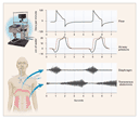
Figure 1. Flow, Airway Pressure, and Inspiratory and Expiratory Muscle
Activity in a Patient with Chronic Obstructive Pulmonary Disease Who
Received Pressure-Support Ventilation at an Airway Pressure of 20 cm of
Water.
The electromyograms in the lower portion of the figure show inspiratory
muscle activity in the patient's diaphragm and expiratory muscle activity in
the transversus abdominis. The patient's increased inspiratory effort caused
the airway pressure to fall below the set sensitivity (2 cm of water), and
inadequate delivery of flow by the ventilator resulted in a scooped contour
on the airway-pressure curve during inspiration. While the ventilator was
still pumping gas into the patient, his expiratory muscles were recruited,
causing a bump in the airway-pressure curve. That the flow never returned to
zero throughout expiration reflected the presence of autopositive
end-expiratory pressure. The broken red line shows airway pressure in
another patient, who generated just enough effort to trigger the ventilator
and in whom there was adequate delivery of gas by the ventilator. Data are
from Jubran et al. 6 <http://content.nejm.org/cgi/content/full/344/26/#R6>
and Parthasarathy et al. 11
<http://content.nejm.org/cgi/content/full/344/26/#R11>
Almost all patients who undergo mechanical ventilation receive some form of
assisted ventilation, with the patient's inspiratory effort triggering the
ventilator. To ensure that the ventilator does not cycle too often, the
clinician sets a threshold for airway pressure that will trigger the
ventilator. This threshold, referred to as set sensitivity, is usually 1
to 2 cm of water. 14 <http://content.nejm.org/cgi/content/full/344/26/#R14>
To reach this threshold, the patient must initiate an inspiratory effort.
But when the threshold is reached, inspiratory neurons do not simply switch
off. Consequently, the patient may expend considerable inspiratory effort
throughout the machine-cycled inflation. 15
<http://content.nejm.org/cgi/content/full/344/26/#R15>
The display of airway pressure and flow tracings on ventilator screens has i
ncreased awareness that inspiratory effort is frequently insufficient to
trigger the ventilator. At high levels of mechanical assistance, up to one
third of a patient's inspiratory efforts may fail to trigger the machine. 9
<http://content.nejm.org/cgi/content/full/344/26/#R9> , 16
<http://content.nejm.org/cgi/content/full/344/26/#R16> , 17
<http://content.nejm.org/cgi/content/full/344/26/#R17> Surprisingly,
unsuccessful triggering is not the result of poor inspiratory effort;
indeed, the effort is more than a third greater when the threshold for
triggering the ventilator is not reached than when it is reached. 9
<http://content.nejm.org/cgi/content/full/344/26/#R9> Breaths that do not
reach the threshold for triggering the ventilator have higher tidal volumes
and shorter expiratory times than do breaths that do trigger the ventilator.
Consequently, elastic-recoil pressure builds up within the thorax in the
form of intrinsic positive end-expiratory pressure (PEEP), or auto-PEEP. 9
<http://content.nejm.org/cgi/content/full/344/26/#R9> To trigger the
ventilator, the patient's inspiratory effort first has to generate a
negative intrathoracic pressure in order to counterbalance the elastic
recoil and then must reach the set sensitivity. The consequences of wasted
inspiratory efforts are not fully known, but they add an unnecessary burden
in patients whose inspiratory muscles are already under stress.
The inspiratory flow rate is initially set at a default value, such as 60
liters per minute. If the delivered flow does not meet the patient's
ventilatory needs, inspiratory effort will increase. 15
<http://content.nejm.org/cgi/content/full/344/26/#R15> Sometimes the flow
is increased in order to shorten the inspiratory time and increase the
expiratory time, especially in patients with inspiratory efforts that are
insufficient to trigger the ventilator. But an increase in flow causes
immediate and persistent tachypnea, and as a result, the expiratory time may
be shortened. 18 <http://content.nejm.org/cgi/content/full/344/26/#R18> In
one study, for example, increases in inspiratory flow from 30 liters per
minute to 60 and 90 liters per minute caused increases in the respiratory
rate of 20 and 41 percent, respectively. 19
<http://content.nejm.org/cgi/content/full/344/26/#R19>
In studies of interactions between the patient's respiratory effort and
mechanical ventilation, remarkably little attention has been paid to the
switch between inspiration and expiration. With the use of pressure-support
ventilation, ventilatory assistance ceases when the patient's inspiratory
flow falls by a preset amount (e.g., to 25 percent of the peak flow). 5
<http://content.nejm.org/cgi/content/full/344/26/#R5> Air flow changes more
slowly in patients with chronic obstructive pulmonary disease than in other
patients, and patients often start to exhale while the ventilator is still
pumping gas into their chests. 6
<http://content.nejm.org/cgi/content/full/344/26/#R6> , 11
<http://content.nejm.org/cgi/content/full/344/26/#R11> In 5 of 12 patients
with chronic obstructive pulmonary disease who were receiving pressure
support of 20 cm of water, expiratory muscles were recruited during
ventilator-induced inflation. 6
<http://content.nejm.org/cgi/content/full/344/26/#R6>
Improving Oxygenation and Preventing Lung Injury
A primary goal of mechanical ventilation is to improve arterial oxygenation.
Improvement is achieved partly through the use of endotracheal intubation to
ensure the delivery of oxygen to the airway and partly through an increase
in airway pressure. Satisfactory oxygenation is easily achieved in most
patients with airway obstruction. The main challenge arises in patients with
alveolar-filling disorders, especially the acute respiratory distress
syndrome a form of noncardiogenic pulmonary edema resulting from severe
acute alveolar injury. It has long been recognized that arterial oxygenation
can be achieved at a lower inspired oxygen concentration by increasing
airway pressure. The goal of using the lowest possible oxygen concentration
to achieve an arterial oxygen saturation of approximately 90 percent has not
changed in decades. What has changed is how this goal is viewed in relation
to other factors, particularly ventilator pressures. In recent years, there
has been a growing tendency to be more concerned about high airway pressures
than about oxygen toxicity, although this shift has been based on a
consensus of opinion rather than on data from studies in patients and
animals.
From the outset, clinicians recognized that mechanical ventilation could
rupture alveoli and cause air leaks. 20
<http://content.nejm.org/cgi/content/full/344/26/#R20> In 1974, Webb and
Tierney showed that mechanical ventilation could also cause ultrastructural
injury, independently of air leaks. 21
<http://content.nejm.org/cgi/content/full/344/26/#R21> Their observations
went largely unnoticed until a decade later, when several investigators
confirmed and extended them. Alveolar overdistention causes changes in
epithelial and endothelial permeability, alveolar hemorrhage, and
hyaline-membrane formation in laboratory animals. 22
<http://content.nejm.org/cgi/content/full/344/26/#R22>
Diffuse infiltrates on chest radiographs originally led clinicians to infer
that lung involvement was homogeneous. But computed tomography (CT) reveals
a patchy pattern: about one third of the lung is unaerated, one third poorly
aerated, and one third normally aerated. 23
<http://content.nejm.org/cgi/content/full/344/26/#R23> , 24
<http://content.nejm.org/cgi/content/full/344/26/#R24> A ventilator-induced
breath will follow the path of least impediment, travelling preferentially
to the normally aerated areas. As a result, these regions are vulnerable to
alveolar overdistention and the type of ventilator-induced lung injury found
in laboratory animals 25
<http://content.nejm.org/cgi/content/full/344/26/#R25> ( Figure 2
<http://content.nejm.org/cgi/content/full/344/26/#F2> ).
<http://content.nejm.org/cgi/content/full/344/26/1986/F2>
View larger version (75K):
[in this window] <http://content.nejm.org/cgi/content/full/344/26/1986/F2>
[in a new window]
<http://content.nejm.org/cgi/content-nw/full/344/26/1986/F2>
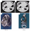
Figure 2. Lung Injury Caused by Mechanical Ventilation in a 31-Year-Old
Woman with the Acute Respiratory Distress Syndrome Due to Amniotic-Fluid
Embolism.
The patient had undergone mechanical ventilation for eight weeks with tidal
volumes of 12 to 15 ml per kilogram of body weight, peak airway pressures of
50 to 70 cm of water, positive end-expiratory pressures of 10 to 15 cm of
water, and a fractional inspired oxygen concentration of 0.80 to 1.00 in
order to achieve a partial pressure of carbon dioxide that was less than 50
mm Hg and a partial pressure of oxygen that was 80 mm Hg or higher. Computed
tomography (CT) performed two days before the patient died revealed a
paramediastinal pneumatocele in the right lung (Panel A, arrowheads) and
numerous intraparenchymal pseudocysts in the left lung (Panel B, black
arrow, open circle, and asterisk).
At autopsy, both lungs were removed and fixed by intrabronchial infusion of
formalin, alcohol, and polyethylene glycol at an insufflation pressure of 30
cm of water. Panel C shows the paramediastinal pneumatocele in the right
lung (arrowheads); the horizontal broken line is the level of the CT
section. Panel D shows a 1-cm-thick section of the left lung, corresponding
to the CT section. Small pseudocysts are present (arrow), and two large
pseudocysts (asterisk and open circle) have compressed and partially
destroyed the parenchyma. The development of these lesions in a patient
without a history of chronic lung disease indicates the harm that may result
with the use of high tidal volumes and airway pressures. The photographs
were kindly provided by Dr. Jean-Jacques Rouby, Hôpital de la
PitiéSalpêtrière, Paris.
A new era of ventilatory management began in 1990, when Hickling et al. 26
<http://content.nejm.org/cgi/content/full/344/26/#R26> reported that
lowering the tidal volume caused a 60 percent decrease in the expected
mortality rate among patients with the acute respiratory distress syndrome.
In a subsequent trial, Amato et al. 27
<http://content.nejm.org/cgi/content/full/344/26/#R27> , 28
<http://content.nejm.org/cgi/content/full/344/26/#R28> randomly assigned
patients to a conventional tidal volume (12 ml per kilogram of body weight)
or to a low tidal volume (less than 6 ml per kilogram). Mortality was
decreased by 46 percent with the lower tidal volume. In a recent study of
861 patients, the Acute Respiratory Distress Syndrome Network 29
<http://content.nejm.org/cgi/content/full/344/26/#R29> confirmed this
benefit: mortality was decreased by 22 percent with a tidal volume of 6 ml
per kilogram as compared with a tidal volume of 12 ml per kilogram. Lowering
the tidal volume, however, failed to improve the outcome in three controlled
trials. 30 <http://content.nejm.org/cgi/content/full/344/26/#R30> , 31
<http://content.nejm.org/cgi/content/full/344/26/#R31> , 32
<http://content.nejm.org/cgi/content/full/344/26/#R32> The discrepant
findings can be explained by differences in trial design. Increased survival
was demonstrable only when the patients undergoing conventional ventilation
had a mean pressure during an end-inspiratory pause (the so-called plateau
pressure, a surrogate for peak alveolar pressure) that exceeded 32 cm of
water. 33 <http://content.nejm.org/cgi/content/full/344/26/#R33>
The pressures pertinent to ventilatory management are the peak inspiratory
pressure, plateau pressure, and end-expiratory pressure. Patients with
airway obstruction may have a very high peak pressure without any increase
in the plateau pressure. Indeed, the gradient between the two is directly
related to the resistance of the airway to airflow. An increase in the peak
inspiratory pressure without a concomitant increase in the plateau pressure
is unlikely to cause alveolar damage. The critical variable is not airway
pressure itself but transpulmonary pressure airway pressure during the
end-inspiratory pause minus pleural pressure. The normal lung is maximally
distended at a transpulmonary pressure between 30 and 35 cm of water, and
higher pressures cause overdistention. Patients with stiff chest walls, such
as those with the acute respiratory distress syndrome due to a nonpulmonary
disorder (e.g., abdominal sepsis), have an elevated pleural pressure. In
such patients, the airway plateau pressure may exceed 35 cm of water without
causing alveolar overdistention.
Clinical decisions based on plateau pressure must take into account the
relation between lung volume and airway pressure in the individual patient.
The pressurevolume curve in patients with the acute respiratory distress
syndrome typically has a sigmoid shape with two discrete bends, called
inflection points ( Figure 3
<http://content.nejm.org/cgi/content/full/344/26/#F3> ). 34
<http://content.nejm.org/cgi/content/full/344/26/#R34> Some investigators
believe that a plateau pressure above the upper bend causes alveolar
overdistention. Reducing the tidal volume lowers the plateau pressure, but
at the cost of hypercapnia. In a study in which 25 patients with the acute
respiratory distress syndrome underwent mechanical ventilation with a tidal
volume of 10 ml per kilogram, 20 had a plateau pressure that was 2 to 14 cm
of water above the upper bend of the pressurevolume curve. 35
<http://content.nejm.org/cgi/content/full/344/26/#R35> Lowering the plateau
pressure to a value that fell below the upper bend required a 22 percent
decrease in the tidal volume, causing the partial pressure of carbon dioxide
to increase from 44 to 77 mm Hg. 35
<http://content.nejm.org/cgi/content/full/344/26/#R35> The partial pressure
of carbon dioxide, in turn, can be decreased by as much as 28 percent by
removing tubing and thus decreasing dead space and increasing the frequency
of ventilator-induced breaths. By virtue of their stiff lungs, patients with
the acute respiratory distress syndrome who do not have an underlying airway
obstruction can tolerate a frequency of 30 breaths per minute without gas
trapping. 36 <http://content.nejm.org/cgi/content/full/344/26/#R36> Severe
hypercapnia can have adverse effects, including increased intracranial
pressure, depressed myocardial contractility, pulmonary hypertension, and
depressed renal blood flow. 37
<http://content.nejm.org/cgi/content/full/344/26/#R37> , 38
<http://content.nejm.org/cgi/content/full/344/26/#R38> The view that these
risks are preferable to the higher plateau pressure required to achieve
normocapnia represents a substantial shift in ventilatory management.
<http://content.nejm.org/cgi/content/full/344/26/1986/F3>
View larger version (26K):
[in this window] <http://content.nejm.org/cgi/content/full/344/26/1986/F3>
[in a new window]
<http://content.nejm.org/cgi/content-nw/full/344/26/1986/F3>
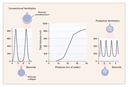
Figure 3. Respiratory PressureVolume Curve and the Effects of Traditional
as Compared with Protective Ventilation in a 70-kg Patient with the Acute
Respiratory Distress Syndrome.
The lower and upper inflection points of the inspiratory pressurevolume
curve (center panel) are at 14 and 26 cm of water, respectively. With
conventional ventilation at a tidal volume of 12 ml per kilogram of body
weight and zero end-expiratory pressure (left-hand panel), alveoli collapse
at the end of expiration. The generation of shear forces during the
subsequent mechanical inflation may tear the alveolar lining, and attaining
an end-inspiratory volume higher than the upper inflection point causes
alveolar overdistention. With protective ventilation at a tidal volume of 6
ml per kilogram (right-hand panel), the end-inspiratory volume remains below
the upper inflection point; the addition of positive end-expiratory pressure
at 2 cm of water above the lower inflection point may prevent alveolar
collapse at the end of expiration and provide protection against the
development of shear forces during mechanical inflation.
Lowering the tidal volume is not without hazards. In addition to the
potential harm of hypercapnia, the volume of aerated lung may be decreased,
39 <http://content.nejm.org/cgi/content/full/344/26/#R39> with a consequent
increase in shunting and worsening oxygenation. One means of minimizing the
loss of lung volume is the use of sighs (i.e., single breaths of large tidal
volume). In one study, increasing the plateau pressure by at least 10 cm of
water during sighs, applied three times a minute over a period of one hour,
caused a 26 percent decrease in shunting, with a 50 percent increase in the
partial pressure of oxygen. 40
<http://content.nejm.org/cgi/content/full/344/26/#R40> It is unknown
whether sighs used at this low frequency cause injury from alveolar
overdistention.
The more usual way of improving oxygenation is through the use of PEEP with
the intention of recruiting previously nonfunctioning lung tissue. Selecting
the right level of PEEP for a given patient with the acute respiratory
distress syndrome is difficult, because the severity of injury varies
throughout the lungs. PEEP can recruit atelectatic areas but may overdistend
normally aerated areas. 41
<http://content.nejm.org/cgi/content/full/344/26/#R41> , 42
<http://content.nejm.org/cgi/content/full/344/26/#R42> In a study involving
six patients with acute lung injury, for example, the use of PEEP at 13 cm
of water resulted in the recruitment of nonaerated portions of lung, with a
gain of 320 ml in volume, but three patients had overdistention of already
aerated portions of lung, with an excess volume of 238 ml. 43
<http://content.nejm.org/cgi/content/full/344/26/#R43>
Overall, about 30 percent of patients with acute lung injury do not benefit
from PEEP or have a fall in the partial pressure of oxygen. 23
<http://content.nejm.org/cgi/content/full/344/26/#R23> , 44
<http://content.nejm.org/cgi/content/full/344/26/#R44> , 45
<http://content.nejm.org/cgi/content/full/344/26/#R45> With the patient in
the supine posture, PEEP generally recruits the regions of the lung closest
to the apex and sternum. 23
<http://content.nejm.org/cgi/content/full/344/26/#R23> Conversely, PEEP can
increase the amount of nonaerated tissue in the regions close to the spine
and the diaphragm. 23 <http://content.nejm.org/cgi/content/full/344/26/#R23>
Among patients in the early stages of the acute respiratory distress
syndrome, those with pulmonary causes, such as pneumonia, are less likely to
benefit from PEEP than are those with nonpulmonary causes, such as
intraabdominal sepsis or extrathoracic trauma. 46
<http://content.nejm.org/cgi/content/full/344/26/#R46> This distinction may
be related to the type of morphologic involvement: pulmonary causes of the
syndrome are characterized by alveolar filling, whereas nonpulmonary causes
are characterized by interstitial edema and alveolar collapse. In the later
stages of the acute respiratory distress syndrome, remodeling and fibrosis
may eliminate this distinction between pulmonary and nonpulmonary causes.
To select the right level of PEEP, some experts recommend bedside
calculation of the pressurevolume curve. With the ventilators currently
used in the United States, calculating the pressurevolume curve is
logistically difficult and technically demanding. 34
<http://content.nejm.org/cgi/content/full/344/26/#R34> Yet many ventilators
have a computer screen, and minor software modifications would make it
feasible to calculate the curve in as little as two minutes as with the
ventilators available in France. 47
<http://content.nejm.org/cgi/content/full/344/26/#R47> Providing this
option on ventilators would increase clinicians' experience with the use of
pressurevolume curves in ventilatory management.
Even if the pressurevolume curve is not calculated at the bedside, it is
useful to select the PEEP level according to this conceptual framework. A
level above the lower bend in the pressurevolume curve is thought to keep
alveoli open at the end of expiration and thus prevent the injury that can
result from shear forces created by the opening and closing of alveoli. 48
<http://content.nejm.org/cgi/content/full/344/26/#R48> , 49
<http://content.nejm.org/cgi/content/full/344/26/#R49> , 50
<http://content.nejm.org/cgi/content/full/344/26/#R50> This level of PEEP
may also prevent an increase in the amount of nonaerated tissue and, thus,
atelectasis. However, the notion that the lower bend signals the level of
PEEP necessary to prevent end-expiratory collapse and that pressures above
the upper bend signal alveolar overdistention is a gross oversimplification.
The relation between the shape of the pressurevolume curve and events at
the alveolar level is confounded by numerous factors and is the subject of
ongoing research and debate. 51
<http://content.nejm.org/cgi/content/full/344/26/#R51> , 52
<http://content.nejm.org/cgi/content/full/344/26/#R52> , 53
<http://content.nejm.org/cgi/content/full/344/26/#R53> , 54
<http://content.nejm.org/cgi/content/full/344/26/#R54> , 55
<http://content.nejm.org/cgi/content/full/344/26/#R55> An understanding of
this relation is also impeded by the difficulty in distinguishing collapsed
lung units from fluid-filled units on CT.
Most patients with the acute respiratory distress syndrome have an increase
in the partial pressure of oxygen when there is a change from the supine to
the prone position. In a study of 16 patients, for example, 12 had an
increase of 9 to 73 mm Hg in the partial pressure of oxygen, and 4 had a
decrease of 7 to 16 mm Hg. 56
<http://content.nejm.org/cgi/content/full/344/26/#R56> The mechanism
responsible for the improvement in the partial pressure of oxygen is not
clear. The attribution of this improvement to lung recruitment has not been
proved. 56 <http://content.nejm.org/cgi/content/full/344/26/#R56> It is now
posited that a prone position causes ventilation to be distributed more
evenly to the various regions of the lungs, 57
<http://content.nejm.org/cgi/content/full/344/26/#R57> , 58
<http://content.nejm.org/cgi/content/full/344/26/#R58> improving the
relation between ventilation and perfusion. 59
<http://content.nejm.org/cgi/content/full/344/26/#R59> , 60
<http://content.nejm.org/cgi/content/full/344/26/#R60>
Discontinuing Mechanical Ventilation
Because mechanical ventilation can have life-threatening complications, it
should be discontinued at the earliest possible time. The process of
discontinuing mechanical ventilation, termed weaning, is one of the most
challenging problems in intensive care, and it accounts for a considerable
proportion of the workload of staff in an intensive care unit. 2
<http://content.nejm.org/cgi/content/full/344/26/#R2>
When mechanical ventilation is discontinued, up to 25 percent of patients
have respiratory distress severe enough to necessitate the reinstitution of
ventilatory support. 61
<http://content.nejm.org/cgi/content/full/344/26/#R61> , 62
<http://content.nejm.org/cgi/content/full/344/26/#R62> Our understanding of
why weaning fails in some patients has advanced considerably in recent
years. Among patients who cannot be weaned, disconnection from the
ventilator is followed almost immediately by an increase in respiratory
frequency and a fall in tidal volume that is, rapid, shallow breathing 63
<http://content.nejm.org/cgi/content/full/344/26/#R63> ( Figure 4
<http://content.nejm.org/cgi/content/full/344/26/#F4> ). As a trial of
spontaneous breathing is continued over the next 30 to 60 minutes, the
respiratory effort increases considerably, reaching more than four times the
normal value at the end of this period. 7
<http://content.nejm.org/cgi/content/full/344/26/#R7> The increased effort
is mainly due to worsening respiratory mechanics. Respiratory resistance
increases progressively over the course of a trial of spontaneous breathing,
reaching about seven times the normal value at the end of the trial; lung
stiffness also increases, reaching five times the normal value; and gas
trapping, measured as auto-PEEP, more than doubles over the course of the
trial. 7 <http://content.nejm.org/cgi/content/full/344/26/#R7> Before
weaning is started, however, the respiratory mechanics in such patients are
similar to those in whom subsequent weaning is successful. 66
<http://content.nejm.org/cgi/content/full/344/26/#R66> Thus, unknown
mechanisms associated with the act of spontaneous breathing cause the
worsening of respiratory mechanics in patients who cannot be weaned from
mechanical ventilation.
<http://content.nejm.org/cgi/content/full/344/26/1986/F4>
View larger version (42K):
[in this window] <http://content.nejm.org/cgi/content/full/344/26/1986/F4>
[in a new window]
<http://content.nejm.org/cgi/content-nw/full/344/26/1986/F4>
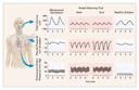
Figure 4. Tidal Volume, Pleural Pressure, and Pulmonary-Artery Pressure in a
Patient Undergoing Assist-Control Ventilation and at the Start and End of a
Failed Trial of Spontaneous Breathing.
During mechanical ventilation, the patient's inspiratory effort is in the
normal range and the pulmonary-artery pressure is 45/22 mm Hg
(systolic/diastolic). At the start of the trial of spontaneous breathing,
the tidal volume falls to 200 ml, the respiratory frequency increases to 33
breaths per minute, there are swings in pleural pressure of 11 cm of water,
and the pulmonary-artery pressure at the end of expiration is 60/28 mm Hg.
At the end of the trial, 45 minutes later, the tidal volume and respiratory
frequency are unchanged, there are swings in pleural pressure of 19 cm of
water, autopositive end-expiratory pressure is 4 cm of water, and the
pulmonary-artery pressure is 60/31 mm Hg. The values in a healthy subject
are tidal volume, 380 ml; respiratory frequency, 17 breaths per minute;
pleural-pressure swings, 3 cm of water; and pulmonary-artery pressure, 18/8
mm Hg. Data are from Tobin et al. 63
<http://content.nejm.org/cgi/content/full/344/26/#R63> , 64
<http://content.nejm.org/cgi/content/full/344/26/#R64> and Jubran et al. 7
<http://content.nejm.org/cgi/content/full/344/26/#R7> , 65
<http://content.nejm.org/cgi/content/full/344/26/#R65>
In addition to the increase in respiratory effort, an unsuccessful attempt
at spontaneous breathing causes considerable cardiovascular stress. 67
<http://content.nejm.org/cgi/content/full/344/26/#R67> Patients can have
substantial increases in right and left ventricular afterload, with
increases of 39 and 27 percent in pulmonary and systemic arterial pressures,
respectively, 64 <http://content.nejm.org/cgi/content/full/344/26/#R64>
most likely because the negative swings in intrathoracic pressure are more
extreme. At the completion of a trial of weaning, the level of oxygen
consumption is equivalent in patients who can be weaned and in those who
cannot. But how the cardiovascular system meets the oxygen demand differs in
the two groups of patients. 64
<http://content.nejm.org/cgi/content/full/344/26/#R64> In those who are
successfully weaned, the oxygen demand is met through an increase in oxygen
delivery, mediated by the expected increase in cardiac output on
discontinuation of positive-pressure ventilation. In patients who cannot be
weaned, the oxygen demand is met through an increase in oxygen extraction,
and these patients have a relative decrease in oxygen delivery. 64
<http://content.nejm.org/cgi/content/full/344/26/#R64> The greater oxygen
extraction causes a substantial decrease in mixed venous oxygen saturation,
contributing to the arterial hypoxemia that occurs in some patients. 64
<http://content.nejm.org/cgi/content/full/344/26/#R64>
Over the course of a trial of spontaneous breathing, about half of patients
in whom the trial fails have an increase in carbon dioxide tension of 10 mm
Hg or more. 7 <http://content.nejm.org/cgi/content/full/344/26/#R7> The
hypercapnia is not usually a consequence of a decrease in minute
ventilation. 63 <http://content.nejm.org/cgi/content/full/344/26/#R63>
Instead, hypercapnia results from rapid, shallow breathing, which causes an
increase in dead-space ventilation. In a small proportion of patients who
cannot be weaned, primary depression of respiratory drive may be responsible
for the hypercapnia. 7 <http://content.nejm.org/cgi/content/full/344/26/#R7>
The discontinuation of mechanical ventilation needs to be carefully timed.
Premature discontinuation places severe stress on the respiratory and
cardiovascular systems, which can impede the patient's recovery. Unnecessary
delays in discontinuation can lead to a host of complications. Decisions
about timing that are based solely on expert clinical judgment are
frequently erroneous. 68
<http://content.nejm.org/cgi/content/full/344/26/#R68> , 69
<http://content.nejm.org/cgi/content/full/344/26/#R69> , 70
<http://content.nejm.org/cgi/content/full/344/26/#R70> Several functional
measures are used to aid decision making. The level of oxygenation must be
satisfactory before one attempts to discontinue mechanical ventilation. Yet
in many patients with satisfactory oxygenation, such attempts fail. The use
of traditional predictors of the success or failure of attempts maximal
inspiratory pressure, vital capacity, and minute ventilation frequently
has false positive or false negative results. 71
<http://content.nejm.org/cgi/content/full/344/26/#R71> A more reliable
predictor is the ratio of respiratory frequency to tidal volume (f/VT). 72
<http://content.nejm.org/cgi/content/full/344/26/#R72> The ratio must be
calculated during spontaneous breathing; calculating it during pressure
support markedly impairs its predictive accuracy. 68
<http://content.nejm.org/cgi/content/full/344/26/#R68> The higher the
ratio, the more severe the rapid, shallow breathing and the greater the
likelihood of unsuccessful weaning. A ratio of 100 best discriminates
between successful and unsuccessful attempts at weaning. In a case of
clinical equipoise that is, a pretest probability of 50 percent an f/VT
of 80, which has a likelihood ratio of 7.5, is associated with almost a 95
percent post-test probability of successful weaning. 73
<http://content.nejm.org/cgi/content/full/344/26/#R73> If the f/VT is
higher than 100, the likelihood ratio is 0.04 and the post-test probability
of successful weaning is less than 5 percent.
Several groups of investigators have evaluated the predictive value of f/VT.
74 <http://content.nejm.org/cgi/content/full/344/26/#R74> , 75
<http://content.nejm.org/cgi/content/full/344/26/#R75> , 76
<http://content.nejm.org/cgi/content/full/344/26/#R76> , 77
<http://content.nejm.org/cgi/content/full/344/26/#R77> , 78
<http://content.nejm.org/cgi/content/full/344/26/#R78> Its positive
predictive value the proportion of patients who are successfully weaned
among those for whom the ratio predicts success has generally been high
(0.8 or higher). The negative predictive value the proportion of patients
who cannot be weaned among those for whom the ratio predicts failure has
sometimes been reported to be low (0.5 or less). Low negative predictive
values have often been reported for patients with a high likelihood of
successful extubation for example, patients undergoing routine
postoperative ventilatory assistance and patients who have tolerated initial
trials of weaning. 75 <http://content.nejm.org/cgi/content/full/344/26/#R75>
, 76 <http://content.nejm.org/cgi/content/full/344/26/#R76>
There are four methods of weaning. 79
<http://content.nejm.org/cgi/content/full/344/26/#R79> The oldest method is
to perform trials of spontaneous breathing several times a day, with the use
of a T-tube circuit containing an enriched supply of oxygen. Initially 5 to
10 minutes in duration, the trials are extended and repeated several times a
day until the patient can sustain spontaneous ventilation for several hours.
This approach has become unpopular because it requires considerable time on
the part of intensive care staff.
The two most common approaches, intermittent mandatory ventilation and
pressure support, decrease ventilatory assistance gradually by respectively
lowering the number of ventilator-assisted breaths or the level of pressure.
When a minimal level of ventilatory assistance can be tolerated, the patient
is extubated. The minimal level of assistance, however, has never been well
defined. For example, pressure support of 6 to 8 cm of water is widely used
to compensate for the resistance imposed by the endotracheal tube and
ventilator circuit. 80
<http://content.nejm.org/cgi/content/full/344/26/#R80> A patient who can
breathe comfortably at this level of pressure support should be able to
tolerate extubation. But if the upper airways are swollen because an
endotracheal tube has been in place for several days, the work engendered by
breathing through the swollen airways is about the same as that caused by
breathing through an endotracheal tube. 81
<http://content.nejm.org/cgi/content/full/344/26/#R81> Accordingly, any
amount of pressure support overcompensates and may give misleading
information about the likelihood that a patient can tolerate extubation.
The fourth method of weaning is to perform a single daily T-tube trial,
lasting for up to two hours. If this trial is successful, the patient is
extubated; if the trial is unsuccessful, the patient is given at least 24
hours of respiratory-muscle rest with full ventilatory support before
another trial is performed. 82
<http://content.nejm.org/cgi/content/full/344/26/#R82>
Until the early 1990s, it was widely believed that all weaning methods were
equally effective, and the physician's judgment was regarded as the critical
determinant. But the results of randomized, controlled trials clearly
indicate that the period of weaning is as much as three times as long with
intermittent mandatory ventilation as with trials of spontaneous breathing.
61 <http://content.nejm.org/cgi/content/full/344/26/#R61> , 62
<http://content.nejm.org/cgi/content/full/344/26/#R62> In a study involving
patients with respiratory difficulties on weaning, trials of spontaneous
breathing halved the weaning time as compared with pressure support 62
<http://content.nejm.org/cgi/content/full/344/26/#R62> ; in another study,
the weaning time was similar with the two methods. 61
<http://content.nejm.org/cgi/content/full/344/26/#R61> Performing trials of
spontaneous breathing once a day is as effective as performing such trials
several times a day 62
<http://content.nejm.org/cgi/content/full/344/26/#R62> but much simpler. In
a recent study, half-hour trials of spontaneous breathing were as effective
as two-hour trials. 83
<http://content.nejm.org/cgi/content/full/344/26/#R83> However, this study
involved all patients being considered for weaning, not just those for whom
there were difficulties with weaning.
A two-stage approach to weaning systematic measurement of predictors,
including f/VT , followed by a single daily trial of spontaneous breathing
was compared with conventional management in a randomized trial. 69
<http://content.nejm.org/cgi/content/full/344/26/#R69> Although the
patients assigned to the two-stage approach were sicker than those assigned
to conventional weaning, they were weaned twice as rapidly. The rate of
complications and the costs of intensive care were also lower with two-stage
management than with conventional management.
When patients can sustain spontaneous ventilation without undue discomfort,
they are extubated. About 10 to 20 percent of such patients require
reintubation. 61 <http://content.nejm.org/cgi/content/full/344/26/#R61> , 62
<http://content.nejm.org/cgi/content/full/344/26/#R62> Mortality among
patients who require reintubation is more than six times as high as
mortality among patients who can tolerate extubation. 83
<http://content.nejm.org/cgi/content/full/344/26/#R83> , 84
<http://content.nejm.org/cgi/content/full/344/26/#R84> The reason for the
higher mortality is unknown; it is not clearly related to the development of
new problems after extubation or to complications of reinserting the tube.
Indeed, the need for reintubation may simply be a marker of a more severe
underlying illness.
In a controlled trial involving patients who could not sustain spontaneous
ventilation, the patients who were extubated and then received noninvasive
ventilation through a face mask had a shorter mean overall period of
ventilatory support (10.2 days) than those who remained intubated and were
weaned by decreasing pressure support (16.6 days). 85
<http://content.nejm.org/cgi/content/full/344/26/#R85> Although this result
is promising, it is not clear how many such patients or which ones could
benefit from this approach.
Other Approaches to Mechanical Ventilation
Noninvasive ventilation, an approach that is becoming more widespread, was
reviewed in the Journal in 1997. 86
<http://content.nejm.org/cgi/content/full/344/26/#R86> Two new approaches
under investigation are liquid ventilation 87
<http://content.nejm.org/cgi/content/full/344/26/#R87> and
proportional-assist ventilation 16
<http://content.nejm.org/cgi/content/full/344/26/#R16> ; they have not yet
been approved for general clinical use.
Conclusions
Since my previous overview of mechanical ventilation in the Journal, we have
gained a better understanding of the pathophysiology associated with
unsuccessful weaning and have learned how to wean patients more efficiently.
We have also learned how ventilator settings influence survival in patients
with the acute respiratory distress syndrome. Less progress has been made in
determining how the ventilator can best be used to achieve maximal
respiratory-muscle rest, which is the most common reason for providing
mechanical ventilation. Although further research may lead to unexpected new
insights, an important challenge for researchers is to identify elements of
our current knowledge that can be incorporated into a clinical management
scheme to improve the outcome for patients who require ventilatory
assistance.
Supported by a Merit Review grant from the Department of Veterans Affairs
Research and Development Service.
I am indebted to Drs. Amal Jubran, Franco Laghi, and Thomas Brack for
helpful criticisms on successive drafts of the manuscript.
Source Information
From the Division of Pulmonary and Critical Care Medicine, Edward Hines,
Jr., Veterans Affairs Hospital and Loyola University of Chicago Stritch
School of Medicine, Hines, Ill.
Address reprint requests to Dr. Tobin at the Division of Pulmonary and
Critical Care Medicine, Edward Hines, Jr., Veterans Affairs Hospital, Rte.
111N, Hines, IL 60141, or at [log in to unmask] <mailto:[log in to unmask]> .
References
1. Tobin MJ. Mechanical ventilation. N Engl J Med 1994;330:1056-1061. [Full
Text]
<http://content.nejm.org/cgi/ijlink?linkType=FULL&journalCode=nejm&resid=330
/15/1056>
2. Esteban A, Anzueto A, Alia I, et al. How is mechanical ventilation
employed in the intensive care unit? An international utilization review. Am
J Respir Crit Care Med 2000;161:1450-1458. [Abstract/Full Text]
<http://content.nejm.org/cgi/ijlink?linkType=ABST&journalCode=ajrccm&resid=1
61/5/1450>
3. Sassoon CSH. Intermittent mandatory ventilation. In: Tobin JM, ed.
Principles and practice of mechanical ventilation. New York: McGraw-Hill,
1994:221-37.
4. Marini JJ, Smith TC, Lamb VJ. External work output and force generation
during synchronized intermittent mechanical ventilation: effect of machine
assistance on breathing effort. Am Rev Respir Dis 1988;138:1169-1179.
[Medline]
<http://content.nejm.org/cgi/external_ref?access_num=3202477&link_type=MED>
5. Brochard L. Pressure support ventilation. In: Tobin MJ, ed. Principles
and practice of mechanical ventilation. New York: McGraw-Hill, 1994:239-57.
6. Jubran A, Van de Graaff WB, Tobin MJ. Variability of patient-ventilator
interaction with pressure support ventilation in patients with chronic
obstructive pulmonary disease. Am J Respir Crit Care Med 1995;152:129-136.
[Abstract]
<http://content.nejm.org/cgi/ijlink?linkType=ABST&journalCode=ajrccm&resid=1
52/1/129>
7. Jubran A, Tobin JM. Pathophysiologic basis of acute respiratory distress
in patients who fail a trial of weaning from mechanical ventilation. Am J
Respir Crit Care Med 1997;155:906-915. [Abstract]
<http://content.nejm.org/cgi/ijlink?linkType=ABST&journalCode=ajrccm&resid=1
55/3/906>
8. Reid WD, Huang J, Bryson S, Walker DC, Belcastro AN. Diaphragm injury and
myofibrillar structure induced by resistive loading. J Appl Physiol
1994;76:176-184. [Medline]
<http://content.nejm.org/cgi/external_ref?access_num=8175503&link_type=MED>
9. Leung P, Jubran A, Tobin MJ. Comparison of assisted ventilator modes on
triggering, patient effort, and dyspnea. Am J Respir Crit Care Med
1997;155:1940-1948. [Abstract]
<http://content.nejm.org/cgi/ijlink?linkType=ABST&journalCode=ajrccm&resid=1
55/6/1940>
10. Anzueto A, Peters JI, Tobin JM, et al. Effects of prolonged controlled
mechanical ventilation on diaphragmatic function in healthy adult baboons.
Crit Care Med 1997;25:1187-1190. [Medline]
<http://content.nejm.org/cgi/external_ref?access_num=9233746&link_type=MED>
11. Parthasarathy S, Jubran A, Tobin MJ. Cycling of inspiratory and
expiratory muscle groups with the ventilator in airflow limitation. Am J
Respir Crit Care Med 1998;158:1471-1478. [Erratum, Am J Respir Crit Care Med
1999;159:1023.] [Abstract/Full Text]
<http://content.nejm.org/cgi/ijlink?linkType=ABST&journalCode=ajrccm&resid=1
58/5/1471>
12. Simon PM, Zurob AS, Wies WM, Leiter JC, Hubmayr RD. Entrainment of
respiration in humans by periodic lung inflations: effect of state and CO2.
Am J Respir Crit Care Med 1999;160:950-960. [Abstract/Full Text]
<http://content.nejm.org/cgi/ijlink?linkType=ABST&journalCode=ajrccm&resid=1
60/3/950>
13. Parthasarathy S, Jubran A, Tobin MJ. Assessment of neural inspiratory
time in ventilator-supported patients. Am J Respir Crit Care Med
2000;162:546-552. [Abstract/Full Text]
<http://content.nejm.org/cgi/ijlink?linkType=ABST&journalCode=ajrccm&resid=1
62/2/546>
14. Sassoon CSH, Gruer SE. Characteristics of the ventilator pressure- and
flow-trigger variables. Intensive Care Med 1995;21:159-168. [Medline]
<http://content.nejm.org/cgi/external_ref?access_num=7775698&link_type=MED>
15. Marini JJ, Capps JS, Culver BH. The inspiratory work of breathing during
assisted mechanical ventilation. Chest 1985;87:612-618. [Abstract]
<http://content.nejm.org/cgi/ijlink?linkType=ABST&journalCode=chest&resid=87
/5/612>
16. Giannouli E, Webster K, Roberts D, Younes M. Response of
ventilator-dependent patients to different levels of pressure support and
proportional assist. Am J Respir Crit Care Med 1999;159:1716-1725.
[Abstract/Full Text]
<http://content.nejm.org/cgi/ijlink?linkType=ABST&journalCode=ajrccm&resid=1
59/6/1716>
17. Sinderby C, Navalesi P, Beck J, et al. Neural control of mechanical
ventilation in respiratory failure. Nat Med 1999;5:1433-1436. [Medline]
<http://content.nejm.org/cgi/external_ref?access_num=10581089&link_type=MED>
18. Puddy A, Younes M. Effect of inspiratory flow rate on respiratory output
in normal subjects. Am Rev Respir Dis 1992;146:787-789. [Medline]
<http://content.nejm.org/cgi/external_ref?access_num=1519864&link_type=MED>
19. Laghi F, Karamchandani K, Tobin MJ. Influence of ventilator settings in
determining respiratory frequency during mechanical ventilation. Am J Respir
Crit Care Med 1999;160:1766-1770. [Abstract/Full Text]
<http://content.nejm.org/cgi/ijlink?linkType=ABST&journalCode=ajrccm&resid=1
60/5/1766>
20. Pierson DJ. Barotrauma and bronchopleural fistula. In: Tobin MJ, ed.
Principles and practice of mechanical ventilation. New York: McGraw-Hill,
1994:813-36.
21. Webb HH, Tierney DF. Experimental pulmonary edema due to intermittent
positive pressure ventilation with high inflation pressures: protection by
positive end-expiratory pressure. Am Rev Respir Dis 1974;110:556-565.
[Medline]
<http://content.nejm.org/cgi/external_ref?access_num=4611290&link_type=MED>
22. Dreyfuss D, Saumon G. Ventilator-induced lung injury: lessons from
experimental studies. Am J Respir Crit Care Med 1998;157:294-323. [Full
Text]
<http://content.nejm.org/cgi/ijlink?linkType=FULL&journalCode=ajrccm&resid=1
57/1/294>
23. Puybasset L, Cluzel P, Chao N, Slutsky AS, Coriat P, Rouby JJ. A
computed tomography scan assessment of regional lung volume in acute lung
injury. Am J Respir Crit Care Med 1998;158:1644-1655. [Abstract/Full Text]
<http://content.nejm.org/cgi/ijlink?linkType=ABST&journalCode=ajrccm&resid=1
58/5/1644>
24. Gattinoni L, Pesenti A, Bombino M, et al. Relationships between lung
computed tomographic density, gas exchange, and PEEP in acute respiratory
failure. Anesthesiology 1988;69:824-832. [Medline]
<http://content.nejm.org/cgi/external_ref?access_num=3057937&link_type=MED>
25. Rouby JJ, Lherm T, Martin de Lassale E, et al. Histologic aspects of
pulmonary barotrauma in critically ill patients with acute respiratory
failure. Intensive Care Med 1993;19:383-389. [Medline]
<http://content.nejm.org/cgi/external_ref?access_num=8270717&link_type=MED>
26. Hickling KG, Henderson SJ, Jackson R. Low mortality associated with low
volume pressure limited ventilation with permissive hypercapnia in severe
adult respiratory distress syndrome. Intensive Care Med 1990;16:372-377.
[Medline]
<http://content.nejm.org/cgi/external_ref?access_num=2246418&link_type=MED>
27. Amato MB, Barbas CS, Medeiros DM, et al. Beneficial effects of the "open
lung approach" with low distending pressures in acute respiratory distress
syndrome: a prospective randomized study on mechanical ventilation. Am J
Respir Crit Care Med 1995;152:1835-1846. [Abstract]
<http://content.nejm.org/cgi/ijlink?linkType=ABST&journalCode=ajrccm&resid=1
52/6/1835>
28. Amato MBP, Barbas CSV, Medeiros DM, et al. Effect of a
protective-ventilation strategy on mortality in the acute respiratory
distress syndrome. N Engl J Med 1998;338:347-354. [Abstract/Full Text]
<http://content.nejm.org/cgi/ijlink?linkType=ABST&journalCode=nejm&resid=338
/6/347>
29. The Acute Respiratory Distress Syndrome Network. Ventilation with lower
tidal volumes as compared with traditional tidal volumes for acute lung
injury and the acute respiratory distress syndrome. N Engl J Med
2000;342:1301-1308. [Abstract/Full Text]
<http://content.nejm.org/cgi/ijlink?linkType=ABST&journalCode=nejm&resid=342
/18/1301>
30. Stewart TE, Meade MO, Cook DJ, et al. Evaluation of a ventilation
strategy to prevent barotrauma in patients at high risk for acute
respiratory distress syndrome. N Engl J Med 1998;338:355-361. [Abstract/Full
Text]
<http://content.nejm.org/cgi/ijlink?linkType=ABST&journalCode=nejm&resid=338
/6/355>
31. Brochard L, Roudot-Thoraval F, Roupie E, et al. Tidal volume reduction
for prevention of ventilator-induced lung injury in acute respiratory
distress syndrome. Am J Respir Crit Care Med 1998;158:1831-1838.
[Abstract/Full Text]
<http://content.nejm.org/cgi/ijlink?linkType=ABST&journalCode=ajrccm&resid=1
58/6/1831>
32. Brower RG, Shanholtz CB, Fessler HE, et al. Prospective, randomized,
controlled clinical trial comparing traditional versus reduced tidal volume
ventilation in acute respiratory distress syndrome patients. Crit Care Med
1999;27:1492-1498. [Medline]
<http://content.nejm.org/cgi/external_ref?access_num=10470755&link_type=MED>
33. Tobin MJ. Culmination of an era in research on the acute respiratory
distress syndrome. N Engl J Med 2000;342:1360-1361. [Full Text]
<http://content.nejm.org/cgi/ijlink?linkType=FULL&journalCode=nejm&resid=342
/18/1360>
34. Brochard L. Respiratory pressure-volume curves. In: Tobin MJ, ed.
Principles and practice of intensive care monitoring. New York: McGraw-Hill,
1998:597-616.
35. Roupie E, Dambrosio M, Servillo G, et al. Titration of tidal volume and
induced hypercapnia in acute respiratory distress syndrome. Am J Respir Crit
Care Med 1995;152:121-128. [Abstract]
<http://content.nejm.org/cgi/ijlink?linkType=ABST&journalCode=ajrccm&resid=1
52/1/121>
36. Richecoeur J, Lu Q, Vieira SR, et al. Expiratory washout versus
optimization of mechanical ventilation during permissive hypercapnia in
patients with severe acute respiratory distress syndrome. Am J Respir Crit
Care Med 1999;160:77-85. [Abstract/Full Text]
<http://content.nejm.org/cgi/ijlink?linkType=ABST&journalCode=ajrccm&resid=1
60/1/77>
37. Feihl F, Perret C. Permissive hypercapnia: how permissive should we be?
Am J Respir Crit Care Med 1994;150:1722-1737. [Medline]
<http://content.nejm.org/cgi/external_ref?access_num=7952641&link_type=MED>
38. Feihl F, Eckert P, Brimioulle S, et al. Permissive hypercapnia impairs
pulmonary gas exchange in the acute respiratory distress syndrome. Am J
Respir Crit Care Med 2000;162:209-215. [Abstract/Full Text]
<http://content.nejm.org/cgi/ijlink?linkType=ABST&journalCode=ajrccm&resid=1
62/1/209>
39. Mead J, Collier C. Relation of volume history of lungs to respiratory
mechanics in anesthetized dogs. J Appl Physiol 1959;14:669-678.
40. Pelosi P, Cadringher P, Bottino N, et al. Sigh in acute respiratory
distress syndrome. Am J Respir Crit Care Med 1999;159:872-880.
[Abstract/Full Text]
<http://content.nejm.org/cgi/ijlink?linkType=ABST&journalCode=ajrccm&resid=1
59/3/872>
41. Dambrosio M, Roupie E, Mollett JJ, et al. Effects of positive
end-expiratory pressure and different tidal volumes on alveolar recruitment
and hyperinflation. Anesthesiology 1997;87:495-503. [Medline]
<http://content.nejm.org/cgi/external_ref?access_num=9316952&link_type=MED>
42. Vieira SR, Puybasset L, Lu Q, et al. A scanographic assessment of
pulmonary morphology in acute lung injury: significance of the lower
inflection point detected on the lung pressure-volume curve. Am J Respir
Crit Care Med 1999;159:1612-1623. [Abstract/Full Text]
<http://content.nejm.org/cgi/ijlink?linkType=ABST&journalCode=ajrccm&resid=1
59/5/1612>
43. Vieira SR, Puybasset L, Richecoeur J, et al. A lung computed tomographic
assessment of positive end-expiratory pressure-induced lung overdistension.
Am J Respir Crit Care Med 1998;158:1571-1577. [Abstract/Full Text]
<http://content.nejm.org/cgi/ijlink?linkType=ABST&journalCode=ajrccm&resid=1
58/5/1571>
44. Horton WG, Cheney FW. Variability of effect of positive end expiratory
pressure. Arch Surg 1975;110:395-398. [Medline]
<http://content.nejm.org/cgi/external_ref?access_num=1096847&link_type=MED>
45. Kanarek DJ, Shannon DC. Adverse effect of positive end-expiratory
pressure on pulmonary perfusion and arterial oxygenation. Am Rev Respir Dis
1975;112:457-459. [Medline]
<http://content.nejm.org/cgi/external_ref?access_num=1099951&link_type=MED>
46. Gattinoni L, Pelosi P, Suter PM, Pedoto A, Vercesi P, Lissoni A. Acute
respiratory distress syndrome caused by pulmonary and extrapulmonary
disease: different syndromes? Am J Respir Crit Care Med 1998;158:3-11.
[Abstract/Full Text]
<http://content.nejm.org/cgi/ijlink?linkType=ABST&journalCode=ajrccm&resid=1
58/1/3>
47. Lu Q, Vieira SR, Richecoeur J, et al. A simple automated method for
measuring pressure-volume curves during mechanical ventilation. Am J Respir
Crit Care Med 1999;159:275-282. [Abstract/Full Text]
<http://content.nejm.org/cgi/ijlink?linkType=ABST&journalCode=ajrccm&resid=1
59/1/275>
48. Argiras EP, Blakeley CR, Dunnill MS, Otremski S, Sykes MK. High PEEP
decreases hyaline membrane formation in surfactant deficient lungs. Br J
Anaesth 1987;59:1278-1285. [Abstract]
<http://content.nejm.org/cgi/ijlink?linkType=ABST&journalCode=brjana&resid=5
9/10/1278>
49. Tremblay L, Valenza F, Ribeiro SP, Li J, Slutsky AS. Injurious
ventilatory strategies increase cytokines and c-fos m-RNA expression in an
isolated rat lung model. J Clin Invest 1997;99:944-952. [Abstract/Full Text]
<http://content.nejm.org/cgi/ijlink?linkType=ABST&journalCode=jci&resid=99/5
/944>
50. Mead J, Takishima T, Leith D. Stress distribution in lungs: a model of
pulmonary elasticity. J Appl Physiol 1970;28:596-608. [Medline]
<http://content.nejm.org/cgi/external_ref?access_num=5442255&link_type=MED>
51. Hickling KG. The pressure-volume curve is greatly modified by
recruitment: a mathematical model of ARDS lungs. Am J Respir Crit Care Med
1998;158:194-202. [Abstract/Full Text]
<http://content.nejm.org/cgi/ijlink?linkType=ABST&journalCode=ajrccm&resid=1
58/1/194>
52. Martynowicz MA, Minor TA, Walters BJ, Hubmayr RD. Regional expansion of
oleic acid-injured lungs. Am J Respir Crit Care Med 1999;160:250-258.
[Abstract/Full Text]
<http://content.nejm.org/cgi/ijlink?linkType=ABST&journalCode=ajrccm&resid=1
60/1/250>
53. Lichtwarck-Aschoff M, Mols G, Hedlund AJ, et al. Compliance is nonlinear
over tidal volume irrespective of positive end-expiratory pressure level in
surfactant-depleted piglets. Am J Respir Crit Care Med 2000;162:2125-2133.
[Abstract/Full Text]
<http://content.nejm.org/cgi/ijlink?linkType=ABST&journalCode=ajrccm&resid=1
62/6/2125>
54. Carney DE, Bredenberg CE, Schiller HJ, et al. The mechanism of lung
volume change during mechanical ventilation. Am J Respir Crit Care Med
1999;160:1697-1702.
55. Harris RS, Hess DR, Venegas JG. An objective analysis of the
pressure-volume curve in the acute respiratory distress syndrome. Am J
Respir Crit Care Med 2000;161:432-439. [Abstract/Full Text]
<http://content.nejm.org/cgi/ijlink?linkType=ABST&journalCode=ajrccm&resid=1
61/2/432>
56. Pelosi P, Tubiolo D, Mascheroni D, et al. Effects of the prone position
on respiratory mechanics and gas exchange during acute lung injury. Am J
Respir Crit Care Med 1998;157:387-393. [Abstract/Full Text]
<http://content.nejm.org/cgi/ijlink?linkType=ABST&journalCode=ajrccm&resid=1
57/2/387>
57. Gattinoni L, Pelosi P, Vitale G, Pesenti A, D'Andrea L, Mascheroni D.
Body position changes redistribute lung computed-tomographic density in
patients with acute respiratory failure. Anesthesiology 1991;74:15-23.
[Medline]
<http://content.nejm.org/cgi/external_ref?access_num=1986640&link_type=MED>
58. Mutoh T, Guest RJ, Lamm WJ, Albert RK. Prone position alters the effect
of volume overload on regional pleural pressures and improves hypoxemia in
pigs in vivo. Am Rev Respir Dis 1992;146:300-306. [Medline]
<http://content.nejm.org/cgi/external_ref?access_num=1489116&link_type=MED>
59. Mure M, Domino KB, Lindahl SG, Hlastala MP, Altemeier WA, Glenny RW.
Regional ventilation-perfusion distribution is more uniform in the prone
position. J Appl Physiol 2000;88:1076-1083. [Abstract/Full Text]
<http://content.nejm.org/cgi/ijlink?linkType=ABST&journalCode=jap&resid=88/3
/1076>
60. Albert RK, Hubmayr RD. The prone position eliminates compression of the
lungs by the heart. Am J Respir Crit Care Med 2000;161:1660-1665.
[Abstract/Full Text]
<http://content.nejm.org/cgi/ijlink?linkType=ABST&journalCode=ajrccm&resid=1
61/5/1660>
61. Brochard L, Rauss A, Benito S, et al. Comparison of three methods of
gradual withdrawing from ventilatory support during weaning from mechanical
ventilation. Am J Respir Crit Care Med 1994;150:896-903. [Abstract]
<http://content.nejm.org/cgi/ijlink?linkType=ABST&journalCode=ajrccm&resid=1
50/4/896>
62. Esteban A, Frutos F, Tobin MJ, et al. A comparison of four methods of
weaning patients from mechanical ventilation. N Engl J Med 1995;332:345-350.
[Abstract/Full Text]
<http://content.nejm.org/cgi/ijlink?linkType=ABST&journalCode=nejm&resid=332
/6/345>
63. Tobin MJ, Perez W, Guenther SM, et al. The pattern of breathing during
successful and unsuccessful trials of weaning from mechanical ventilation.
Am Rev Respir Dis 1986;134:1111-1118. [Medline]
<http://content.nejm.org/cgi/external_ref?access_num=3789513&link_type=MED>
64. Tobin MJ, Chadha TS, Jenouri G, Birch SJ, Gazeroglu HB, Sackner MA.
Breaking patterns. 1. Normal subjects. Chest 1983;84:202-205. [Abstract]
<http://content.nejm.org/cgi/ijlink?linkType=ABST&journalCode=chest&resid=84
/2/202>
65. Jubran A, Mathru M, Dries D, Tobin MJ. Continuous recordings of mixed
venous oxygen saturation during weaning from mechanical ventilation and the
ramifications thereof. Am J Respir Crit Care Med 1998;158:1763-1769.
[Abstract/Full Text]
<http://content.nejm.org/cgi/ijlink?linkType=ABST&journalCode=ajrccm&resid=1
58/6/1763>
66. Jubran A, Tobin MJ. Passive mechanics of lung and chest wall in patients
who failed or succeeded in trials of weaning. Am J Respir Crit Care Med
1997;155:916-921. [Abstract]
<http://content.nejm.org/cgi/ijlink?linkType=ABST&journalCode=ajrccm&resid=1
55/3/916>
67. Lemaire F, Teboul JL, Cinotti L, et al. Acute left ventricular
dysfunction during unsuccessful weaning from mechanical ventilation.
Anesthesiology 1988;69:171-179. [Medline]
<http://content.nejm.org/cgi/external_ref?access_num=3044189&link_type=MED>
68. Stroetz RW, Hubmayr RD. Tidal volume maintenance during weaning with
pressure support. Am J Respir Crit Care Med 1995;152:1034-1040. [Abstract]
<http://content.nejm.org/cgi/ijlink?linkType=ABST&journalCode=ajrccm&resid=1
52/3/1034>
69. Ely EW, Baker AM, Dunagan DP, et al. Effect on the duration of
mechanical ventilation of identifying patients capable of breathing
spontaneously. N Engl J Med 1996;335:1864-1869. [Abstract/Full Text]
<http://content.nejm.org/cgi/ijlink?linkType=ABST&journalCode=nejm&resid=335
/25/1864>
70. Coplin WM, Pierson DJ, Cooley KD, Newell DW, Rubenfeld GD. Implications
of extubation delay in brain-injured patients meeting standard weaning
criteria. Am J Respir Crit Care Med 2000;161:1530-1536. [Abstract/Full Text]
<http://content.nejm.org/cgi/ijlink?linkType=ABST&journalCode=ajrccm&resid=1
61/5/1530>
71. Tobin MJ, Alex CG. Discontinuation of mechanical ventilation. In: Tobin
MJ, ed. Principles and practice of mechanical ventilation. New York:
McGraw-Hill, 1994:1177-206.
72. Yang KL, Tobin MJ. A prospective study of indexes predicting the outcome
of trials of weaning from mechanical ventilation. N Engl J Med
1991;324:1445-1450. [Abstract]
<http://content.nejm.org/cgi/ijlink?linkType=ABST&journalCode=nejm&resid=324
/21/1445>
73. Jaeschke RZ, Meade MO, Guyatt GH, Keenan SP, Cook DJ. How to use
diagnostic test articles in the intensive care unit: diagnosing weanability
using f/Vt. Crit Care Med 1997;25:1514-1521. [Medline]
<http://content.nejm.org/cgi/external_ref?access_num=9295825&link_type=MED>
74. Sassoon CSH, Mahutte CK. Airway occlusion pressure and breathing pattern
as predictors of weaning outcome. Am Rev Respir Dis 1993;148:860-866.
[Medline]
<http://content.nejm.org/cgi/external_ref?access_num=8214939&link_type=MED>
75. Epstein SK. Etiology of extubation failure and the predictive value of
the rapid shallow breathing index. Am J Respir Crit Care Med
1995;152:545-549. [Abstract]
<http://content.nejm.org/cgi/ijlink?linkType=ABST&journalCode=ajrccm&resid=1
52/2/545>
76. Jacob B, Chatila W, Manthous CA. The unassisted respiratory rate/tidal
volume ratio accurately predicts weaning outcome in postoperative patients.
Crit Care Med 1997;25:253-257. [Medline]
<http://content.nejm.org/cgi/external_ref?access_num=9034260&link_type=MED>
77. Vallverdu I, Calaf N, Subirana M, Net A, Benito S, Mancebo J. Clinical
characteristics, respiratory functional parameters, and outcome of a
two-hour T-piece trial in patients weaning from mechanical ventilation. Am J
Respir Crit Care Med 1998;158:1855-1862. [Abstract/Full Text]
<http://content.nejm.org/cgi/ijlink?linkType=ABST&journalCode=ajrccm&resid=1
58/6/1855>
78. Maldonado A, Bauer TT, Ferrer M, et al. Capnometric recirculation gas
tonometry and weaning from mechanical ventilation. Am J Respir Crit Care Med
2000;161:171-176. [Abstract/Full Text]
<http://content.nejm.org/cgi/ijlink?linkType=ABST&journalCode=ajrccm&resid=1
61/1/171>
79. Tobin MJ. 1999 Donald F Egan Scientific Lecture: weaning from mechanical
ventilation: what have we learned? Respir Care 2000;45:417-431. [Medline]
<http://content.nejm.org/cgi/external_ref?access_num=10780038&link_type=MED>
80. Brochard L, Rua F, Lorino H, Lemaire F, Harf A. Inspiratory pressure
support compensates for the additional work of breathing caused by the
endotracheal tube. Anesthesiology 1991;75:739-745. [Medline]
<http://content.nejm.org/cgi/external_ref?access_num=1952198&link_type=MED>
81. Straus C, Louis B, Isabey D, Lemaire F, Harf A, Brochard L. Contribution
of the endotracheal tube and the upper airway to breathing workload. Am J
Respir Crit Care Med 1998;157:23-30. [Abstract/Full Text]
<http://content.nejm.org/cgi/ijlink?linkType=ABST&journalCode=ajrccm&resid=1
57/1/23>
82. Laghi F, D'Alfonso N, Tobin MJ. Pattern of recovery from diaphragmatic
fatigue over 24 hours. J Appl Physiol 1995;79:539-546. [Medline]
<http://content.nejm.org/cgi/external_ref?access_num=7592215&link_type=MED>
83. Esteban A, Alia I, Tobin MJ, et al. Effect of spontaneous breathing
trial duration on outcome of attempts to discontinue mechanical ventilation.
Am J Respir Crit Care Med 1999;159:512-518. [Abstract/Full Text]
<http://content.nejm.org/cgi/ijlink?linkType=ABST&journalCode=ajrccm&resid=1
59/2/512>
84. Epstein SK, Ciubotaru RL, Wong JB. Effect of failed extubation on the
outcome of mechanical ventilation. Chest 1997;112:186-192. [Abstract]
<http://content.nejm.org/cgi/ijlink?linkType=ABST&journalCode=chest&resid=11
2/1/186>
85. Nava S, Ambrosino N, Clini E, et al. Noninvasive mechanical ventilation
in the weaning of patients with respiratory failure due to chronic
obstructive pulmonary disease: a randomized, controlled trial. Ann Intern
Med 1998;128:721-728. [Medline]
<http://content.nejm.org/cgi/external_ref?access_num=9556465&link_type=MED>
86. Hillberg RE, Johnson DC. Noninvasive ventilation. N Engl J Med
1997;337:1746-1752. [Full Text]
<http://content.nejm.org/cgi/ijlink?linkType=FULL&journalCode=nejm&resid=337
/24/1746>
87. Leach CL, Greenspan JS, Rubenstein SD, et al. Partial liquid ventilation
with perflubron in premature infants with severe respiratory distress
syndrome. N Engl J Med 1996;335:761-767. [Abstract/Full Text]
<http://content.nejm.org/cgi/ijlink?linkType=ABST&journalCode=nejm&resid=335
/11/761>
Edward E. Rylander, M.D.
Diplomat American Board of Family Practice.
Diplomat American Board of Palliative Medicine.
|


