The New England Journal of Medicine
Original Article
Volume 345:1863-1869
December 27, 2001
Number 26
Coronary Magnetic Resonance Angiography for the Detection of Coronary
Stenoses
W. Yong Kim, M.D., Ph.D., Peter G. Danias, M.D., Ph.D., Matthias Stuber,
Ph.D., Scott D. Flamm, M.D., Sven Plein, M.D., Eike Nagel, M.D., Susan E.
Langerak, M.Sc., Oliver M. Weber, Ph.D., Erik M. Pedersen, M.D., Ph.D.,
Matthias Schmidt, M.D., René M. Botnar, Ph.D., and Warren J. Manning, M.D.
ABSTRACT
Background An accurate, noninvasive technique for the diagnosis of coronary
disease would be an important advance. We investigated the accuracy of
coronary magnetic resonance angiography among patients with suspected
coronary disease in a prospective, multicenter study.
Methods Coronary magnetic resonance angiography was performed during free
breathing in 109 patients before elective x-ray coronary angiography, and
the results of the two diagnostic procedures were compared.
Results A total of 636 of 759 proximal and middle segments of coronary
arteries (84 percent) were interpretable on magnetic resonance angiography.
In these segments, 78 (83 percent) of 94 clinically significant lesions
(those with a >=50 percent reduction in diameter on x-ray angiography) were
also detected by magnetic resonance angiography. Overall, coronary magnetic
resonance angiography had an accuracy of 72 percent (95 percent confidence
interval, 63 to 81 percent) in diagnosing coronary artery disease. The
sensitivity, specificity, and accuracy for patients with disease of the left
main coronary artery or three-vessel disease were 100 percent (95 percent
confidence interval, 97 to 100 percent), 85 percent (95 percent confidence
interval, 78 to 92 percent), and 87 percent (95 percent confidence interval,
81 to 93 percent), respectively. The negative predictive values for any
coronary artery disease and for left main artery or three-vessel disease
were 81 percent (95 percent confidence interval, 73 to 89 percent) and 100
percent (95 percent confidence interval, 97 to 100 percent), respectively.
Conclusions Among patients referred for their first x-ray coronary
angiogram, three-dimensional coronary magnetic resonance angiography allows
for the accurate detection of coronary artery disease of the proximal and
middle segments. This noninvasive approach reliably identifies (or rules
out) left main coronary artery or three-vessel disease.
_____
Despite progress in prevention and early diagnosis, coronary artery disease
remains the leading cause of death in both men and women in the United
States 1 <http://content.nejm.org/cgi/content/full/345/26/#R1> and
throughout the Western world. Invasive x-ray coronary angiography remains
the gold standard for the identification of clinically significant coronary
artery disease. Although numerous noninvasive tests have been developed to
assist in the identification of patients with coronary artery disease, a
substantial minority of patients referred for elective diagnostic x-ray
coronary angiography are found not to have clinically significant coronary
stenosis (defined as a reduction in the luminal diameter of at least 50
percent). 2 <http://content.nejm.org/cgi/content/full/345/26/#R2> A
noninvasive test that could directly assess the integrity of the coronary
lumen would therefore be desirable.
Coronary magnetic resonance angiography makes possible the noninvasive
visualization of the major epicardial coronary arteries in the majority of
subjects. Since the first reports by Paulin et al. 3
<http://content.nejm.org/cgi/content/full/345/26/#R3> and Edelman et al., 4
<http://content.nejm.org/cgi/content/full/345/26/#R4> coronary magnetic
resonance angiography has undergone technological advances leading to
enhanced spatial resolution and the possibility of imaging while the patient
is breathing freely, but assessment of its usefulness has been hampered by
the lack of standardized hardware, software, and scanning protocols. The
results from single-center studies therefore vary considerably. 5
<http://content.nejm.org/cgi/content/full/345/26/#R5> , 6
<http://content.nejm.org/cgi/content/full/345/26/#R6> , 7
<http://content.nejm.org/cgi/content/full/345/26/#R7> , 8
<http://content.nejm.org/cgi/content/full/345/26/#R8> , 9
<http://content.nejm.org/cgi/content/full/345/26/#R9> , 10
<http://content.nejm.org/cgi/content/full/345/26/#R10> , 11
<http://content.nejm.org/cgi/content/full/345/26/#R11> , 12
<http://content.nejm.org/cgi/content/full/345/26/#R12> , 13
<http://content.nejm.org/cgi/content/full/345/26/#R13> , 14
<http://content.nejm.org/cgi/content/full/345/26/#R14> Coronary magnetic
resonance angiography 15
<http://content.nejm.org/cgi/content/full/345/26/#R15> , 16
<http://content.nejm.org/cgi/content/full/345/26/#R16> performed while the
patient is breathing freely has reached sufficient technical maturity to
allow more widespread application with a standardized protocol. Therefore,
we conducted a prospective, international, multicenter study to determine
the clinical usefulness of coronary magnetic resonance angiography in the
diagnosis of native-vessel coronary artery disease.
Methods
Participating Institutions
The subjects were recruited from seven institutions: Skejby Hospital, Aarhus
University Hospital, Aarhus, Denmark (7 subjects), the German Heart
Institute, Berlin, Germany (18 subjects), Beth Israel Deaconess Medical
Center, Boston (18 subjects), St. Luke's Episcopal Hospital, Houston (25
subjects), Leeds General Infirmary, Leeds, United Kingdom (23 subjects),
Leiden University Medical Center, Leiden, the Netherlands (9 subjects), and
the University and Eidgenoessische Technische Hochschule Zurich, Zurich,
Switzerland (9 subjects). The review board at each institution approved the
study, and written informed consent was obtained from all subjects. In all
cases, coronary magnetic resonance angiography was performed before x-ray
angiography.
Patients
The study population consisted of 109 subjects who were consecutively
enrolled between June 24, 1999, and October 18, 2000. The subjects could be
of either sex and had to be at least 21 years of age with sinus rhythm and
with a body weight of 100 kg or less and to be scheduled to undergo elective
x-ray coronary angiography for suspected coronary artery disease within 14
days. The exclusion criteria were a contraindication to magnetic resonance
imaging 17 <http://content.nejm.org/cgi/content/full/345/26/#R17> (for
example, a pacemaker, intraauricular implants, or intracranial clips),
previous x-ray coronary angiography or thoracotomy, claustrophobia,
orthopnea, and inability to take sublingual nitroglycerin (as a result, for
example, of aortic stenosis or obstructive cardiomyopathy).
Protocol for Magnetic Resonance Angiography
Each center performed coronary magnetic resonance angiography according to a
standard protocol 16 <http://content.nejm.org/cgi/content/full/345/26/#R16>
with use of common hardware and software on a 1.5-T system (Gyroscan ACS-NT,
Philips Medical Systems, Best, the Netherlands) equipped with PowerTrak 6000
gradients (23 mT per meter, 219 µsec rise time). The subjects were examined
with a commercial five-element cardiac synergy receiver coil during
uncoached free breathing. To compensate for artifacts due to respiratory
motion, a right hemidiaphragmatic navigator 18
<http://content.nejm.org/cgi/content/full/345/26/#R18> with real-time slice
correction 19 <http://content.nejm.org/cgi/content/full/345/26/#R19> and a
5-mm end-expiratory gating window were used. For cardiac synchronization and
monitoring, three or four 20
<http://content.nejm.org/cgi/content/full/345/26/#R20> electrodes were
placed on the left anterior hemithorax of the subject with the R wave of the
electrocardiogram used as a trigger for image acquisition. All coronary
images were acquired in mid-diastole. 19
<http://content.nejm.org/cgi/content/full/345/26/#R19>
Magnetic Resonance Localization Scans
The first magnetic resonance localizing scan (approximately one minute)
employed a multistack and multislice, segmented k-space gradientecho
sequence for localization of the heart and diaphragm in three orthogonal
planes (transverse, sagittal, and coronal). 15
<http://content.nejm.org/cgi/content/full/345/26/#R15> , 16
<http://content.nejm.org/cgi/content/full/345/26/#R16> From the coronal
data set, a navigator-gated transverse three-dimensional segmented echo
localizing planar scan (approximately two minutes) with 40 slices was
acquired around the base of the heart to cover the region extending from the
apex of the left ventricle to the pulmonary artery. 16
<http://content.nejm.org/cgi/content/full/345/26/#R16> This allowed
identification of the course of the major right and left coronary arteries.
With the use of a three-point planscan tool, 16
<http://content.nejm.org/cgi/content/full/345/26/#R16> a plane through the
major axis of the proximal and middle segments of the right coronary artery
was subsequently prescribed.
Three-Dimensional Magnetic Resonance Angiography
Coronary magnetic resonance angiography was performed, after sublingual
administration of isosorbide dinitrate (2.5 mg), with the use of a
three-dimensional segmented k-space gradientecho sequence (echo time, 2.2
msec; repetition time, 7.7 msec) during free breathing (for about 10 to 15
minutes). 15 <http://content.nejm.org/cgi/content/full/345/26/#R15> , 16
<http://content.nejm.org/cgi/content/full/345/26/#R16> For contrast
enhancement between blood and the surrounding myocardium and epicardial fat,
a T2-weighted preparation prepulse and a frequency-selective fat-saturation
prepulse were applied. 15
<http://content.nejm.org/cgi/content/full/345/26/#R15> For the right
coronary artery, a double-oblique three-dimensional volume was imaged with
use of the coordinates prescribed by the three-point planscan tool. For the
left main, left anterior descending, and left circumflex coronary arteries,
a double-oblique transverse three-dimensional volume with anteriorposterior
and leftright angulations (5 degrees each) was imaged with the volume
centered on the origin of the left main coronary artery (as defined from the
second localizing scan). For both the left and the right coronary systems,
the three-dimensional volumes were reconstructed to 20 slices, with an
individual slice thickness of 1.5 mm. A field of view of 360 mm and a
512-by-360 matrix yielded an in-plane voxel size of 0.7 by 1.0 mm. No signal
averaging was performed.
Analysis of Coronary Magnetic Resonance Angiograms
Source coronary magnetic resonance angiograms were evaluated at each site
(before x-ray angiography) and again by consensus of two experienced
investigators (from the core magnetic resonance imaging laboratory at the
Beth Israel Deaconess Medical Center) who were blinded to the patients'
clinical data and the x-ray data. The original source images were analyzed
by scrolling through individual slices from the three-dimensional data set
with the use of a commercial software package (EasyVision 4.0, Philips
Medical Systems, Best, the Netherlands). Seven coronary segments were
evaluated: the left main coronary artery and the proximal and middle
segments of the left anterior descending coronary artery (0 to 2 cm and 2 to
4 cm), the left circumflex coronary artery (0 to 1.5 cm and 1.5 to 3 cm),
and the right coronary artery (0 to 2 cm and 2 to 5 cm). For each segment,
image quality was visually graded 21
<http://content.nejm.org/cgi/content/full/345/26/#R21> as 1, indicating
poor or uninterpretable (coronary artery visible, with markedly blurred
borders or edges); 2, good (coronary artery visible, with moderately blurred
borders or edges); 3, very good (coronary artery visible, with mildly
blurred borders or edges); or 4, excellent (coronary artery visible, with
sharply defined borders or edges). If the segments were not imaged or if the
image quality was graded as poor or uninterpretable (grade 1), no further
evaluation was performed. Segments that were not visualized or that were
graded as poor or uninterpretable were not included in the subsequent
analysis. Images of good, very good, and excellent quality (grades 2, 3, and
4) were further classified according to the visual assessment of the
coronary-artery lumen as having no coronary artery disease, minimal disease,
or clinically significant disease (if there was prominent attenuation of the
coronary-lumen signal).
Acquisition and Analysis of Coronary X-Ray Angiograms
Conventional x-ray coronary angiography was performed by standard techniques
22 <http://content.nejm.org/cgi/content/full/345/26/#R22> and in multiple
projections after sublingual administration of isosorbide dinitrate (2.5
mg). An experienced invasive cardiologist at the institution where
angiography was performed analyzed each x-ray angiogram without knowledge of
the magnetic resonance data. Each coronary vessel was assessed, and the
visual estimation of the segment and the maximal percentage reduction of the
luminal diameter for each lesion were reported. In addition, in 99 patients
(91 percent), quantitative x-ray angiographic analysis was performed by an
independent core laboratory (Brigham and Women's Hospital Angiographic Core
Laboratory, Boston) without access to the magnetic resonance data.
Quantitative angiographic analysis was performed according to a standard
algorithm, 23 <http://content.nejm.org/cgi/content/full/345/26/#R23> with
clinically significant disease defined as stenosis of at least 50 percent of
the vessel diameter. For 10 subjects for whom quantitative analysis was not
performed because of administrative issues (e.g., images could not be
located or were unreadable), the visual evaluation reported from the site
was used for the analyses.
Statistical Analysis
The data were retained at the Beth Israel Deaconess Medical Center, and the
primary data analysis was performed by one of us. For each individual vessel
and for each patient, the sensitivity, specificity, and accuracy (percentage
of segments correctly classified) were determined, as well as the positive
and negative predictive values for coronary magnetic resonance angiography
as compared with x-ray coronary angiography. All data are reported as means
±SD or rates with 95 percent confidence intervals.
Results
All subjects completed coronary magnetic resonance angiography without
complications. Of the 109 subjects, 69 percent were men ( Table 1
<http://content.nejm.org/cgi/content/full/345/26/#T1> ). The majority of the
subjects had a history of chest pain, smoking, and hypercholesterolemia.
Sixty-four of the subjects (59 percent) had x-ray angiographic evidence of
coronary artery disease, including 12 percent with three-vessel disease
Table 1 <http://content.nejm.org/cgi/content/full/345/26/#T1> ).
View this table:
[in this window] <http://content.nejm.org/cgi/content/full/345/26/1863/T1>
[in a new window]
<http://content.nejm.org/cgi/content-nw/full/345/26/1863/T1>
Table 1. Demographic, Clinical, and X-Ray Angiographic Characteristics of
the 109 Study Patients.
The mean total magnetic resonance scanning time (including scout imaging)
was 70 minutes (median, 66; range, 33 to 145). The median interval between
the performance of coronary magnetic resonance angiography and x-ray
angiography was 1 day (mean, 3; range, 0 to 14), with no clinical cardiac
events reported between the examinations. Right coronary x-ray angiography
was not performed in two subjects. Thus, 759 coronary segments were
potentially available for analysis. Of these, 636 (84 percent) could be
assessed by coronary magnetic resonance angiography; the proportions of
segments for which images could be assessed ranged from 68 percent (for the
middle left circumflex coronary artery) to 93 percent (for the proximal and
middle right coronary artery) ( Table 2
<http://content.nejm.org/cgi/content/full/345/26/#T2> ). Coronary segments
were not interpretable by magnetic resonance angiography when scans were not
obtained because of time constraints (39 segments), when image quality was
poor (grade 1, 58 segments), or when the three-dimensional volume did not
include the segment (26 segments).
View this table:
[in this window] <http://content.nejm.org/cgi/content/full/345/26/1863/T2>
[in a new window]
<http://content.nejm.org/cgi/content-nw/full/345/26/1863/T2>
Table 2. Number of Interpretable Coronary-Artery Segments and Quality of the
Image on Coronary Magnetic Resonance Angiography.
In the magnetic resonance angiographic assessment of diagnostic accuracy for
each individual vessel, we included all vessels for which the proximal
segment was graded as having an image quality of at least 2 (good or
better). In the analyses for any coronary artery disease and for left main
coronary artery or three-vessel disease, 103 subjects (94 percent) were
eligible according to the consensus readings, and 101 (93 percent) were
eligible according to the readings at the sites. Patients were considered
eligible if clinically significant coronary artery disease identified on
x-ray angiography was found in any coronary artery segment with a readable
magnetic resonance image.
The overall image quality was 2.6±0.7 ( Table 2
<http://content.nejm.org/cgi/content/full/345/26/#T2> ). Individual segment
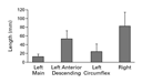
lengths are shown in Figure 1
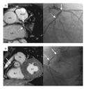
<http://content.nejm.org/cgi/content/full/345/26/#F1> . An example of a
coronary magnetic resonance angiogram and a corresponding x-ray contrast
angiogram for a patient with left and right coronary artery disease are
shown in Figure 2 <http://content.nejm.org/cgi/content/full/345/26/#F2> .
<http://content.nejm.org/cgi/content/full/345/26/1863/F1>
View larger version (4K):
[in this window] <http://content.nejm.org/cgi/content/full/345/26/1863/F1>
[in a new window]
<http://content.nejm.org/cgi/content-nw/full/345/26/1863/F1>
Figure 1. Mean (+SD) Lengths of Segments of Coronary Vessels Visualized by
Coronary Magnetic Resonance Angiography.
<http://content.nejm.org/cgi/content/full/345/26/1863/F2>
View larger version (114K):
[in this window] <http://content.nejm.org/cgi/content/full/345/26/1863/F2>
[in a new window]
<http://content.nejm.org/cgi/content-nw/full/345/26/1863/F2>
Figure 2. Coronary Angiography in a 53-Year-Old Man with Exertional Chest
Pain.
Panel A shows a coronary magnetic resonance angiogram (left) and a
corresponding x-ray coronary angiogram (right) indicating a severe lesion at
the bifurcation of the left anterior descending coronary artery and the left
circumflex coronary artery, involving the left main coronary artery (solid
arrows), and a more distal focal stenosis of the left circumflex coronary
artery (broken arrows). Panel B shows a coronary magnetic resonance
angiogram (left) and a corresponding x-ray angiogram (right) indicating two
stenoses of the proximal (solid arrows) and middle (broken arrows) right
coronary artery. AA denotes ascending aorta, LA left atrium, RVOT right
ventricular outflow tract, PA pulmonary artery, RV right ventricle, and LV
left ventricle.
Seventy-eight of 94 clinically significant coronary stenoses (83 percent)
identified on x-ray angiography were correctly identified on magnetic
resonance angiography. The sensitivity of coronary magnetic resonance
angiography for identifying a patient as having clinically significant
coronary artery disease, according to the consensus of the two interpreters,
was 93 percent (95 percent confidence interval, 88 to 98 percent) ( Table 3
<http://content.nejm.org/cgi/content/full/345/26/#T3> ). All four patients
who had clinically significant coronary artery disease that was not
diagnosed by coronary magnetic resonance angiography had isolated
single-vessel disease, with two (50 percent) having isolated left circumflex
artery disease. In the consensus interpretation, the overall diagnostic
accuracy of coronary magnetic resonance angiography in identifying a patient
as having any coronary artery disease was 72 percent (95 percent confidence
interval, 63 to 81 percent), increasing to 87 percent (95 percent confidence
interval, 81 to 93 percent) for the identification of a patient with left
main coronary artery or three-vessel disease. The prevalence, sensitivity,
specificity, and positive and negative predictive values for individual
coronary vessels and patients according to the consensus and the
site-reported interpretations are summarized in Table 3
<http://content.nejm.org/cgi/content/full/345/26/#T3> .
View this table:
[in this window] <http://content.nejm.org/cgi/content/full/345/26/1863/T3>
[in a new window]
<http://content.nejm.org/cgi/content-nw/full/345/26/1863/T3>
Table 3. Diagnostic Accuracy of Coronary Magnetic Resonance Angiography to
Detect Stenoses of >=50 Percent.
Discussion
In this prospective, multicenter study comparing noncontrast coronary
magnetic resonance angiography with x-ray angiography among patients
referred for a first elective coronary angiogram, we found that coronary
magnetic resonance angiography had a high sensitivity, negative predictive
value, and overall accuracy for detecting coronary artery disease,
especially in subjects with left main coronary artery disease or
three-vessel disease. Coronary magnetic resonance angiography is not
exercise-dependent, and its high negative predictive value suggests that it
may have a role in ruling out clinically significant coronary disease in
this population of patients, among whom the prevalence of disease is
intermediate. Indeed, 41 percent of study subjects had no clinically
significant coronary artery disease, a prevalence similar to that in
previously published data, 2
<http://content.nejm.org/cgi/content/full/345/26/#R2> a fact that
emphasizes the need for an accurate, noninvasive technique that can rule out
clinically significant disease before invasive x-ray coronary angiography.
On the basis of the finding of no clinically significant disease on magnetic
resonance angiography, x-ray angiography could have been avoided in 18
subjects (according to the consensus reading) or 25 subjects (according to
the site reading) those with true negative results ( Table 3
<http://content.nejm.org/cgi/content/full/345/26/#T3> ), or 42 to 58 percent
of subjects without clinically significant coronary artery disease. With the
use of standardized technology and a standardized scanning protocol, all
patients with left main coronary artery or three-vessel disease were
identified as having clinically significant coronary artery disease. These
data therefore support the use of coronary magnetic resonance angiography to
identify (or rule out) left main coronary artery disease or three-vessel
disease reliably. Such information is clinically relevant, since surgical
revascularization in patients with such disease is associated with a more
favorable long-term survival benefit. 24
<http://content.nejm.org/cgi/content/full/345/26/#R24>
The protocol for three-dimensional, noncontrast, free-breathing coronary
magnetic resonance angiography facilitated visualization of the vast
majority of the proximal and middle segments of the left main, left anterior
descending, and right coronary arteries. Coronary magnetic resonance
angiography would detect 94 percent of all patients with any coronary artery
disease or with left main coronary artery or three-vessel disease. The left
circumflex artery was less reliably visualized, but isolated disease of this
artery was found in only 4 percent of subjects, suggesting that the absence
of clinically significant disease in the remaining coronary system makes
left circumflex artery disease unlikely. This observation is in agreement
with other reports. 25
<http://content.nejm.org/cgi/content/full/345/26/#R25> The accuracy of
coronary magnetic resonance angiography for the detection of coronary
disease in the left circumflex artery was also low in prior single-center
studies. 5 <http://content.nejm.org/cgi/content/full/345/26/#R5> , 6
<http://content.nejm.org/cgi/content/full/345/26/#R6> , 9
<http://content.nejm.org/cgi/content/full/345/26/#R9> , 13
<http://content.nejm.org/cgi/content/full/345/26/#R13> This poor accuracy
may be due to the relatively small caliber and posterior location of the
circumflex artery, which results in a lower signal-to-noise ratio because of
the increased distance from the artery to the receiver coils.
Other minimally invasive imaging methods have recently been advocated for
coronary-artery imaging. Electron-beam computed tomography is a highly
sensitive technique for detecting calcium in the coronary arteries. 2
<http://content.nejm.org/cgi/content/full/345/26/#R2> Recent studies using
multislice computed tomography in combination with iodinated contrast medium
to visualize the coronary-artery lumen demonstrated very good diagnostic
accuracy for detecting coronary artery disease when image quality was
adequate (that is, when 70 to 80 percent of images could be assessed). 26
<http://content.nejm.org/cgi/content/full/345/26/#R26> , 27
<http://content.nejm.org/cgi/content/full/345/26/#R27> , 28
<http://content.nejm.org/cgi/content/full/345/26/#R28> A potential
advantage of this method is the acquisition of a complete data set during a
single, though prolonged, breath-holding period (30 to 40 seconds). As
compared with computed tomography, the magnetic resonance approach has the
advantage of requiring no exposure to ionizing radiation or injection of a
contrast agent, and it allows for more comfortable free breathing during the
entire examination. Both the magnetic resonance 29
<http://content.nejm.org/cgi/content/full/345/26/#R29> and computed
tomographic approaches are safe in patients with intracoronary stents, but
interpretation is difficult.
Coronary magnetic resonance angiography has already been demonstrated to be
of clinical value for the assessment of anomalous coronary artery disease,
and it is often superior to x-ray coronary angiography in delineating the
course of the anomalous vessels. 30
<http://content.nejm.org/cgi/content/full/345/26/#R30> , 31
<http://content.nejm.org/cgi/content/full/345/26/#R31> , 32
<http://content.nejm.org/cgi/content/full/345/26/#R32> , 33
<http://content.nejm.org/cgi/content/full/345/26/#R33> However, coronary
magnetic resonance angiography was considered an investigational technique
for the assessment of stenotic native-vessel disease in task-force reports
from Europe and the United States in 1998. 34
<http://content.nejm.org/cgi/content/full/345/26/#R34> , 35
<http://content.nejm.org/cgi/content/full/345/26/#R35> The results of
single-center investigations of coronary magnetic resonance angiography that
used different hardware, software, and scanning protocols have been
variable. 5 <http://content.nejm.org/cgi/content/full/345/26/#R5> , 6
<http://content.nejm.org/cgi/content/full/345/26/#R6> , 7
<http://content.nejm.org/cgi/content/full/345/26/#R7> , 8
<http://content.nejm.org/cgi/content/full/345/26/#R8> , 9
<http://content.nejm.org/cgi/content/full/345/26/#R9> , 10
<http://content.nejm.org/cgi/content/full/345/26/#R10> , 11
<http://content.nejm.org/cgi/content/full/345/26/#R11> , 12
<http://content.nejm.org/cgi/content/full/345/26/#R12> , 13
<http://content.nejm.org/cgi/content/full/345/26/#R13> , 14
<http://content.nejm.org/cgi/content/full/345/26/#R14> Single-center
experience (often including patients who had previously undergone
angiography or a coronary intervention) may also be difficult to translate
into general clinical practice. Clinical acceptance of coronary magnetic
resonance angiography will probably require standardization to ensure
optimal test results. The findings of the present multicenter study should
reflect the clinical value of coronary magnetic resonance angiography more
accurately, because we evaluated a relatively large number of patients at
seven international institutions and used common hardware and software and a
common scanning protocol. Only one of the seven participating institutions
had extensive experience with coronary magnetic resonance angiography.
Furthermore, the independent consensus analyses and those reported from
individual sites were quite similar ( Table 3
<http://content.nejm.org/cgi/content/full/345/26/#T3> ).
Subgroups of patients who may initially benefit from coronary magnetic
resonance angiography are likely to include patients who present with severe
left ventricular systolic dysfunction in the absence of a clinical history
of myocardial infarction. For these patients, the underlying disease process
is either severe multivessel coronary artery disease or nonischemic
cardiomyopathy. Conventional stress tests are often inaccurate in this
group, resulting in frequent referral for diagnostic coronary angiography.
Although this possibility was not directly tested in our study, the data
suggest that coronary magnetic resonance angiography may be able to
discriminate between these two causes; thus, x-ray angiography could be
avoided for those without magnetic resonance evidence of coronary disease.
It should be remembered, however, that coronary magnetic resonance
angiography was unable to assess 16 percent of coronary segments and that 6
percent of the study patients could not be assessed for the presence of any
coronary disease or for left main coronary artery or three-vessel disease.
<http://weeklybriefings.org/feature.asp?strXmlDoc=3452601>
Supported in part by Philips Medical Systems, Best, the Netherlands, which
funded the quantitative coronary angiographic analysis (but not the
selection of the analysis laboratory or data interpretation) and by an
Established Investigatorship Grant from the American Heart Association,
Dallas (9740003N, to Dr. Manning).
Source Information
From the Cardiovascular Division, Department of Medicine (W.Y.K., P.G.D., M.
Stuber, R.M.B., W.J.M.), and the Department of Radiology (W.J.M.), Beth
Israel Deaconess Medical Center and Harvard Medical School, Boston; the
Magnetic Resonance Center, Department of Cardiology, and Institute of
Experimental Clinical Research, Skejby Hospital, Aarhus University Hospital,
Aarhus, Denmark (W.Y.K., E.M.P.); Philips Medical Systems, Best, the
Netherlands (M. Stuber, R.M.B.); St. Luke's Episcopal Hospital and the Texas
Heart Institute, Houston (S.D.F.); the Yorkshire Heart Centre, Leeds General
Infirmary, Leeds, United Kingdom (S.P.); Internal Medicine Cardiology,
German Heart Institute, Berlin, Germany (E.N.); the Department of Radiology
and Cardiology, Leiden University Medical Center, Leiden, the Netherlands
(S.E.L.); the Institute for Biomedical Engineering, University of Zurich,
and the Swiss Federal Institute of Technology Zurich, Zurich, Switzerland
(O.M.W.); and the Klinik und Poliklinik für Nuklearmedizin der Universität
zu Köln, Cologne, Germany (M. Schmidt).
Address reprint requests to Dr. Manning at the Beth Israel Deaconess Medical
Center, 330 Brookline Ave., Boston, MA 02215, or at
[log in to unmask] <mailto:[log in to unmask]> .
References
1. 2001 Heart and stroke statistical update. Dallas: American Heart
Association, 2000.
2. Budoff MJ, Georgiou D, Brody A, et al. Ultrafast computed tomography as a
diagnostic modality in the detection of coronary artery disease: a
multicenter study. Circulation 1996;93:898-904. [Abstract/Full Text]
<http://content.nejm.org/cgi/ijlink?linkType=ABST&journalCode=circulationaha
&resid=93/5/898>
3. Paulin S, von Schulthess GK, Fossel E, Krayenbuehl HP. MR imaging of the
aortic root and proximal coronary arteries. AJR Am J Roentgenol
1987;148:665-670. [Medline]
<http://content.nejm.org/cgi/external_ref?access_num=3493645&link_type=MED>
4. Edelman RR, Manning WJ, Burstein D, Paulin S. Coronary arteries:
breath-hold MR angiography. Radiology 1991;181:641-643. [Abstract]
<http://content.nejm.org/cgi/ijlink?linkType=ABST&journalCode=radiology&resi
d=181/3/641>
5. Manning WJ, Li W, Edelman RR. A preliminary report comparing magnetic
resonance coronary angiography with conventional angiography. N Engl J Med
1993;328:828-832. [Erratum, N Engl J Med 1993;330:152.] [Abstract/Full Text]
<http://content.nejm.org/cgi/ijlink?linkType=ABST&journalCode=nejm&resid=328
/12/828>
6. Duerinckx AJ, Urman MK. Two-dimensional coronary MR angiography: analysis
of initial clinical results. Radiology 1994;193:731-738. [Abstract]
<http://content.nejm.org/cgi/ijlink?linkType=ABST&journalCode=radiology&resi
d=193/3/731>
7. Pennell DJ, Bogren HG, Keegan J, Firmin DN, Underwood SR. Assessment of
coronary artery stenosis by magnetic resonance imaging. Heart
1996;75:127-133. [Abstract]
<http://content.nejm.org/cgi/ijlink?linkType=ABST&journalCode=heartjnl&resid
=75/2/127>
8. Oshinski JN, Hofland L, Mukundan S Jr, Dixon WT, Parks WJ, Pettigrew RI.
Two-dimensional coronary MR angiography without breath holding. Radiology
1996;201:737-743. [Abstract]
<http://content.nejm.org/cgi/ijlink?linkType=ABST&journalCode=radiology&resi
d=201/3/737>
9. Post JC, van Rossum AC, Hofman MB, de Cock CC, Valk J, Visser CA.
Clinical utility of two-dimensional magnetic resonance angiography in
detecting coronary artery disease. Eur Heart J 1997;18:426-433. [Medline]
<http://content.nejm.org/cgi/external_ref?access_num=9076379&link_type=MED>
10. Müller MF, Fleisch M, Kroeker R, Chatterjee T, Meier B, Vock P. Proximal
coronary artery stenosis: three-dimensional MRI with fat saturation and
navigator echo. J Magn Reson Imaging 1997;7:644-651. [Medline]
<http://content.nejm.org/cgi/external_ref?access_num=9243382&link_type=MED>
11. Woodard PK, Li D, Haacke EM, et al. Detection of coronary stenoses on
source and projection images using three-dimensional MR angiography with
retrospective respiratory gating: preliminary experience. AJR Am J
Roentgenol 1998;170:883-888. [Abstract]
<http://content.nejm.org/cgi/ijlink?linkType=ABST&journalCode=ajronline&resi
d=170/4/883>
12. van Geuns RJ, Wielopolski PA, de Bruin HG, et al. MR coronary
angiography with breath-hold targeted volumes: preliminary clinical results.
Radiology 2000;217:270-277. [Abstract/Full Text]
<http://content.nejm.org/cgi/ijlink?linkType=ABST&journalCode=radiology&resi
d=217/1/270>
13. Regenfus M, Ropers D, Achenbach S, et al. Noninvasive detection of
coronary artery stenosis using contrast-enhanced three-dimensional
breath-hold magnetic resonance coronary angiography. J Am Coll Cardiol
2000;36:44-50. [Medline]
<http://content.nejm.org/cgi/external_ref?access_num=10898411&link_type=MED>
14. Moustapha AI, Pereyra M, Muthupillai R, Wilson JM, Flamm SD. Coronary
magnetic resonance angiography using a free-breathing T2 weighted,
three-dimensional gradient echo sequence with navigator respiratory and ECG
gating can be used to detect coronary artery disease. J Am Coll Cardiol
2001;37:Suppl A:380A-380A.abstract
15. Botnar RM, Stuber M, Danias PG, Kissinger KV, Manning WJ. Improved
coronary artery definition with T2-weighted, free-breathing
three-dimensional coronary MRA. Circulation 1999;99:3139-3148.
[Abstract/Full Text]
<http://content.nejm.org/cgi/ijlink?linkType=ABST&journalCode=circulationaha
&resid=99/24/3139>
16. Stuber M, Botnar RM, Danias PG, et al. Double-oblique free-breathing
high resolution three-dimensional coronary magnetic resonance angiography. J
Am Coll Cardiol 1999;34:524-531. [Medline]
<http://content.nejm.org/cgi/external_ref?access_num=10440168&link_type=MED>
17. Sawyer-Glover AM, Shellock FG. Pre-MRI procedure screening:
recommendations and safety considerations for biomedical implants and
devices. J Magn Reson Imaging 2000;12:92-106. [Erratum, J Magn Reson Imaging
2000;12:510.] [Medline]
<http://content.nejm.org/cgi/external_ref?access_num=10931569&link_type=MED>
18. Ehman RL, Felmlee JP. Adaptive technique for high-definition MR imaging
of moving structures. Radiology 1989;173:255-263. [Abstract]
<http://content.nejm.org/cgi/ijlink?linkType=ABST&journalCode=radiology&resi
d=173/1/255>
19. Stuber M, Botnar RM, Danias PG, Kissinger KV, Manning WJ. Submillimeter
three-dimensional coronary MR angiography with real-time navigator
correction: comparison of navigator locations. Radiology 1999;212:579-587.
[Abstract/Full Text]
<http://content.nejm.org/cgi/ijlink?linkType=ABST&journalCode=radiology&resi
d=212/2/579>
20. Fischer SE, Wickline SA, Lorenz CH. Novel real-time R-wave detection
algorithm based on the vectorcardiogram for accurate gated magnetic
resonance acquisitions. Magn Reson Med 1999;42:361-370. [Medline]
<http://content.nejm.org/cgi/external_ref?access_num=10440961&link_type=MED>
21. McConnell MV, Khasgiwala VC, Savord BJ, et al. Comparison of respiratory
suppression methods and navigator locations for MR coronary angiography. AJR
Am J Roentgenol 1997;168:1369-1375. [Abstract]
<http://content.nejm.org/cgi/ijlink?linkType=ABST&journalCode=ajronline&resi
d=168/5/1369>
22. Baim DS, Grossman W, eds. Cardiac catheterization, angiography, and
intervention. 5th ed. Philadelphia: Williams & Wilkins, 1996.
23. van der Zwet PM, Reiber JH. A new approach for the quantification of
complex lesion morphology: the gradient field transform: basic principles
and validation results. J Am Coll Cardiol 1994;24:216-224. [Medline]
<http://content.nejm.org/cgi/external_ref?access_num=8006269&link_type=MED>
24. Eagle KA, Guyton RA, Davidoff R, et al. ACC/AHA guidelines for coronary
artery bypass graft surgery: executive summary and recommendations: a report
of the American College of Cardiology/American Heart Association Task Force
on Practice Guidelines (Committee to Revise the 1991 Guidelines for Coronary
Artery Bypass Graft Surgery). Circulation 1999;100:1464-1480. [Full Text]
<http://content.nejm.org/cgi/ijlink?linkType=FULL&journalCode=circulationaha
&resid=100/13/1464>
25. Hillis LD, Winniford MD. Frequency of severe (70% or more) narrowing of
the right, left anterior descending, and left circumflex coronary arteries
in right dominant circulations with coronary artery disease. Am J Cardiol
1987;59:358-359. [Medline]
<http://content.nejm.org/cgi/external_ref?access_num=3812289&link_type=MED>
26. Rensing BJ, Bongaerts A, van Geuns RJ, van Ooijen P, Oudkerk M, de
Feyter PJ. Intravenous coronary angiography by electron beam computed
tomography: a clinical evaluation. Circulation 1998;98:2509-2512.
[Abstract/Full Text]
<http://content.nejm.org/cgi/ijlink?linkType=ABST&journalCode=circulationaha
&resid=98/23/2509>
27. Achenbach S, Moshage W, Ropers D, Nossen J, Daniel WG. Value of
electron-beam computed tomography for the noninvasive detection of
high-grade coronary-artery stenoses and occlusions. N Engl J Med
1998;339:1964-1971. [Abstract/Full Text]
<http://content.nejm.org/cgi/ijlink?linkType=ABST&journalCode=nejm&resid=339
/27/1964>
28. Nieman K, Oudkerk M, Rensing BJ, et al. Coronary angiography with
multi-slice computed tomography. Lancet 2001;357:599-603. [Medline]
<http://content.nejm.org/cgi/external_ref?access_num=11558487&link_type=MED>
29. Hug J, Nagel E, Bornstedt A, Schnackenburg B, Oswald H, Fleck E.
Coronary arterial stents: safety and artifacts during MR imaging. Radiology
2000;216:781-787. [Abstract/Full Text]
<http://content.nejm.org/cgi/ijlink?linkType=ABST&journalCode=radiology&resi
d=216/3/781>
30. McConnell MV, Ganz P, Selwyn AP, Li W, Edelman RR, Manning WJ.
Identification of anomalous coronary arteries and their anatomic course by
magnetic resonance coronary angiography. Circulation 1995;92:3158-3162.
[Abstract/Full Text]
<http://content.nejm.org/cgi/ijlink?linkType=ABST&journalCode=circulationaha
&resid=92/11/3158>
31. Post JC, van Rossum AC, Bronzwaer JG, et al. Magnetic resonance
angiography of anomalous coronary arteries: a new gold standard for
delineating the proximal course? Circulation 1995;92:3163-3171.
[Abstract/Full Text]
<http://content.nejm.org/cgi/ijlink?linkType=ABST&journalCode=circulationaha
&resid=92/11/3163>
32. Taylor AM, Thorne SA, Rubens MB, et al. Coronary artery imaging in grown
up congenital heart disease: complementary role of magnetic resonance and
x-ray coronary angiography. Circulation 2000;101:1670-1678. [Abstract/Full
Text]
<http://content.nejm.org/cgi/ijlink?linkType=ABST&journalCode=circulationaha
&resid=101/14/1670>
33. Razmi RM, Meduri A, Chun W, Rathi VK, Pohost GM. Coronary magnetic
resonance angiography (CMRA): the gold standard for determining the proximal
course of anomalous coronary arteries. J Am Coll Cardiol 2001;37:Suppl
A:380A-380A.abstract
34. Budinger TF, Berson A, McVeigh ER, et al. Cardiac MR imaging: report of
a working group sponsored by the National Heart, Lung, and Blood Institute.
Radiology 1998;208:573-576. [Medline]
<http://content.nejm.org/cgi/external_ref?access_num=9722831&link_type=MED>
35. Task Force of the European Society of Cardiology, Association of
European Paediatric Cardiologists. The clinical role of magnetic resonance
in cardiovascular disease. Eur Heart J 1998;19:19-39. [Medline]
<http://content.nejm.org/cgi/external_ref?access_num=9503174&link_type=MED>
Appendix
The following investigators also participated in this study: A.D. Blankholm,
Skejby Hospital, Aarhus University Hospital, Aarhus, Denmark; C. Klein, E.
Fleck, J. Hug, and A. Bornstedt, German Heart Institute, Berlin, Germany;
K.V. Kissinger and L.A. Goepfert, Beth Israel Deaconess Medical Center,
Boston; A. Moustapha, M. Pereyra, B. Lambert, J.M. Wilson, and R.
Muthupillai, St. Luke's Episcopal Hospital, Houston; M.U. Sivananthan, J.P
Ridgway, T.R. Jones, and T.N. Bloomer, Leeds General Infirmary, Leeds,
United Kingdom; A. de Roos, P. Kunz, H. Lamb, J.W. Jukema, and E.E. van der
Wall, Leiden University Medical Center, Leiden, the Netherlands; and J.
Schwitter, University Hospital, Zurich, Switzerland.
Edward E. Rylander, M.D.
Diplomat American Board of Family Practice.
Diplomat American Board of Palliative Medicine.
|


