The New England Journal of Medicine
Original Article
Volume 347:5-12
July 4, 2002
Number 1
Widespread Coronary Inflammation in Unstable Angina
Antonino Buffon, M.D., Luigi M. Biasucci, M.D., Giovanna Liuzzo, M.D.,
Giuseppe D'Onofrio, M.D., Filippo Crea, M.D., and Attilio Maseri, M.D.
ABSTRACT
Background Inflammation within vulnerable coronary plaques may cause
unstable angina by promoting rupture and erosion. In unstable angina,
activated leukocytes may be found in peripheral and coronary-sinus blood,
but it is unclear whether they are selectively activated in the vascular bed
of the culprit stenosis.
Methods We measured the content neutrophil myeloperoxidase content in the
cardiac and femoral circulations in five groups of patients: two groups with
unstable angina and stenosis in either the left anterior descending coronary
artery (24 patients) or the right coronary artery (9 patients); 13 with
chronic stable angina; 13 with variant angina and recurrent ischemia; and 6
controls. Blood samples were taken from the aorta, the femoral vein, and the
great cardiac vein, which selectively drains blood from the left but not the
right coronary artery.
Results The neutrophil myeloperoxidase content of aortic blood was similar
in both groups of patients with unstable angina (3.9 and 5.5, with
negative values representing depletion of the enzyme due to neutrophil
activation) and significantly lower than in the other three groups (P<0.05).
Independently of the site of the stenosis, the neutrophil myeloperoxidase
content in blood from the great cardiac vein was significantly decreased in
both groups of patients with unstable angina (6.4 in those with a left
coronary lesion and 6.6 in those with a right coronary lesion), but not in
patients with stable angina and multiple stenoses, patients with variant
angina and recurrent ischemia, or controls. There was also a significant
transcoronary reduction in myeloperoxidase content in both groups with
unstable angina.
Conclusions The widespread activation of neutrophils across the coronary
vascular bed in patients with unstable angina, regardless of the location of
the culprit stenosis, challenges the concept of a single vulnerable plaque
in unstable coronary syndromes.
_____
The hypothesis that inflammation of a vulnerable plaque is responsible for
the development of acute coronary syndromes 1
<http://content.nejm.org/cgi/content/full/347/1/#R1> , 2
<http://content.nejm.org/cgi/content/full/347/1/#R2> , 3
<http://content.nejm.org/cgi/content/full/347/1/#R3> , 4
<http://content.nejm.org/cgi/content/full/347/1/#R4> , 5
<http://content.nejm.org/cgi/content/full/347/1/#R5> is stimulating a
variety of techniques for the detection and stabilization of vulnerable
plaques. 6 <http://content.nejm.org/cgi/content/full/347/1/#R6> , 7
<http://content.nejm.org/cgi/content/full/347/1/#R7> , 8
<http://content.nejm.org/cgi/content/full/347/1/#R8> , 9
<http://content.nejm.org/cgi/content/full/347/1/#R9> , 10
<http://content.nejm.org/cgi/content/full/347/1/#R10> Yet, it is unclear
whether the inflammatory process is confined to a single vulnerable plaque
or whether it is more widespread in the coronary vasculature.
The possibility of widespread inflammation of the coronary arterial bed is
suggested by the recent report of multiple complex coronary plaques in
patients with acute myocardial infarction 11
<http://content.nejm.org/cgi/content/full/347/1/#R11> and by previous
postmortem findings of multiple fresh thrombi in patients with unstable
angina 12 <http://content.nejm.org/cgi/content/full/347/1/#R12> and of
multiple fissured, thrombosed plaques. 13
<http://content.nejm.org/cgi/content/full/347/1/#R13> , 14
<http://content.nejm.org/cgi/content/full/347/1/#R14> A widespread acute
inflammatory process in the coronary arterial bed would have important
implications for a clearer understanding of the pathogenesis, and eventually
for the treatment and prevention, of acute coronary syndromes. By
"widespread," we mean involvement of more than one major coronary artery. By
measuring leukocyte expression of CD11b and CD18 in aortic and
coronary-sinus blood, Mazzone et al. 15
<http://content.nejm.org/cgi/content/full/347/1/#R15> and de Servi et al.
16 <http://content.nejm.org/cgi/content/full/347/1/#R16> demonstrated a
transcoronary inflammatory activation of monocytes and neutrophils in
patients with unstable angina. Such activation was not detectable in aortic
blood. Unfortunately, these authors did not assess the correlation between
activation and the location of the culprit coronary stenosis responsible for
the angina. 15 <http://content.nejm.org/cgi/content/full/347/1/#R15> , 16
<http://content.nejm.org/cgi/content/full/347/1/#R16> Marked activation of
neutrophils was also detected in the peripheral blood of patients with
unstable angina, but not in those with stable angina or in controls.
Activation was detected by measuring the neutrophil myeloperoxidase content,
which is an index of more advanced inflammatory activation than that
identified by measuring CD11b and CD18 expression. 17
<http://content.nejm.org/cgi/content/full/347/1/#R17> , 18
<http://content.nejm.org/cgi/content/full/347/1/#R18>
We ascertained whether the activation of neutrophils, presumably due to
inflammation, in patients with unstable angina was confined to the vascular
bed perfused by the vessel with the culprit coronary stenosis, or whether it
also involved the vascular bed of angiographically normal or nearly normal
arteries. We selected patients with coronary stenoses of either the left
anterior descending or the right coronary artery. We simultaneously measured
the neutrophil myeloperoxidase content in blood from the aorta, the femoral
vein, and the great cardiac vein, which selectively drains blood from the
left anterior descending coronary artery but not the right coronary artery.
19 <http://content.nejm.org/cgi/content/full/347/1/#R19> Patients with
stable angina and stenosis of the left anterior descending coronary artery,
patients with variant angina and recurrent ischemia of the left anterior
descending coronary artery, and patients without coronary disease (controls)
were also studied.
Methods
Patients
We studied a total of 65 patients, divided into five groups. Two of the
groups consisted of the 33 patients who had Braunwald class IIIB unstable
angina. Coronary angiography showed that the coronary stenosis responsible
for the angina (the culprit stenosis) was in the left anterior descending
coronary artery in 24 of these patients (the first group), and in the right
coronary artery in the other 9 patients (the second group). The remaining
three groups were made up of 13 patients with chronic stable angina and
stenosis in the left anterior descending coronary artery; 13 patients with
active variant angina and recurrent spasm in the left anterior descending
coronary artery, which was documented by testing with ergonovine; and 6
control patients with mild mitral stenosis, atrial septal defect, or
supraventricular tachycardia and a normal coronary angiogram.
Patients with a recent myocardial infarction (within three months), prior
coronary interventions, an occluded coronary vessel, a culprit coronary
stenosis in the circumflex branch, or intercurrent infective or inflammatory
disorders were excluded from the study. No patients were taking
antiinflammatory agents other than aspirin (up to 100 mg daily).
The protocol was approved by the ethics committee of the Catholic University
of Rome, and all patients gave written informed consent.
Protocol
Serum levels of C-reactive protein were measured on admission and used as a
marker of systemic inflammation. Cardiac catheterization was performed
within a mean (±SD) of 2±1 days. Before the injection of a contrast agent,
all patients underwent sampling of blood from the right femoral vein and
simultaneous sampling of blood from the aorta and great cardiac vein for the
measurement of neutrophil myeloperoxidase. In both groups of patients with
unstable angina, in order to demonstrate that the great cardiac vein
selectively drained blood from the left anterior descending but not from the
right coronary artery, the blood oxygen saturation in the great cardiac vein
was determined before and after the injection of 1.0 mg of isosorbide
dinitrate into the left anterior descending or the right coronary artery,
according to the location of the culprit stenosis. The venousarterial
differences in neutrophil and leukocyte counts through the coronary and
peripheral circulations were also determined.
The myeloperoxidase content was determined by using a hematologic analyzer
(Bayer H*1), which measures the differential leukocyte count as well as the
cell count by automated flow cytochemistry, as previously described. 17
<http://content.nejm.org/cgi/content/full/347/1/#R17> The H*1 computer
software calculates a myeloperoxidase index of the mean myeloperoxidase
content in the neutrophil population. In healthy subjects, this index is
close to 0. Positive values characterize neutrophils rich in
myeloperoxidase, and negative values characterize neutrophils depleted of
myeloperoxidase as a consequence of their activation. A lower
myeloperoxidase index in blood from the great cardiac vein or the femoral
vein, as compared with the aorta, was taken as an index of neutrophil
activation through the coronary or femoral vascular bed. C-reactive protein
levels were measured by a high-sensitivity, latex-enhanced
immunonephelometric assay (Dade Behring BN II analyzer). 20
<http://content.nejm.org/cgi/content/full/347/1/#R20> The working range of
the assay was 0.175 to 1100 mg per liter, and the coefficient of variation
was less than 5 percent.
Statistical Analysis
Because the myeloperoxidase index did not have a normal distribution,
nonparametric tests were used: the MannWhitney test and the KruskalWallis
test with multiple-comparison procedures (Dunn's method) for comparisons
between groups, and the Friedman test and the Wilcoxon test with the
Bonferroni correction for comparisons within groups. Correlations were
determined with use of Spearman's rank-correlation coefficient. 21
<http://content.nejm.org/cgi/content/full/347/1/#R21> The leukocyte and
neutrophil counts had a normal distribution and were evaluated by analysis
of variance for repeated measures with the Bonferroni correction. Chi-square
statistics were used for categorical variables. A P value of less than 0.05
(two-tailed) was considered to indicate statistical significance. Data are
reported as medians and ranges or as means ±SD, as appropriate.
Results
The demographic, clinical, and angiographic characteristics of the patients
are reported in Table 1 <http://content.nejm.org/cgi/content/full/347/1/#T1>
and Table 2 <http://content.nejm.org/cgi/content/full/347/1/#T2> . Anginal
symptoms before coronary angiography were similar in patients who had
unstable angina with a left coronary lesion, those who had unstable angina
with a right coronary lesion, and those who had variant angina ( Table 1
<http://content.nejm.org/cgi/content/full/347/1/#T1> ).
View this table:
[in this window] <http://content.nejm.org/cgi/content/full/347/1/5/T1>
[in a new window] <http://content.nejm.org/cgi/content-nw/full/347/1/5/T1>
Table 1. Clinical Characteristics of the Study Patients.
View this table:
[in this window] <http://content.nejm.org/cgi/content/full/347/1/5/T2>
[in a new window] <http://content.nejm.org/cgi/content-nw/full/347/1/5/T2>
Table 2. Pattern of Coronary Disease.
The blood oxygen saturation in the great cardiac vein markedly increased
after injection of isosorbide dinitrate (1 mg) into the left anterior
descending coronary artery of patients who had unstable angina with a left
coronary lesion, but not after injection into the right coronary artery of
patients who had unstable angina with a right coronary lesion (P=0.001 by
two-way analysis of variance). The median change in the blood oxygen
saturation as a result of the isosorbide dinitrate injection differed
significantly between the two groups (52.4 percent vs. 12.2 percent,
P=0.04), indicating that positioning the catheter in the great cardiac vein
allowed for selective sampling of the blood draining from the vascular bed
of the left anterior descending coronary artery. The leukocyte and
neutrophil counts in the aorta, great cardiac vein, and femoral vein were
similar; no differences were observed among groups ( Table 1
<http://content.nejm.org/cgi/content/full/347/1/#T1> ). Among patients who
had unstable angina with a right coronary lesion, the territory of the left
anterior descending coronary artery had no wall irregularities in three
patients, wall irregularities alone in three patients, and stenosis of 30 to
50 percent of the luminal diameter in the remaining three patients.
Therefore, the atherosclerotic involvement was much smaller than that
observed in patients who had unstable angina with a left coronary lesion and
those who had chronic stable angina ( Table 2
<http://content.nejm.org/cgi/content/full/347/1/#T2> ). 22
<http://content.nejm.org/cgi/content/full/347/1/#R22>
Neutrophil Activation in the Systemic Circulation
The median aortic myeloperoxidase indexes did not differ significantly
between patients who had unstable angina with a left coronary lesion (3.9)
and those who had unstable angina with a right coronary lesion (5.5,
P=0.21), but they were significantly lower than those observed in patients
with stable angina (+0.1), patients with variant angina (+0.1), and controls
(0.8) (P<0.05 for all comparisons). The ranges for all values are reported
in Table 1 <http://content.nejm.org/cgi/content/full/347/1/#T1> and
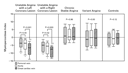
illustrated in Figure 1 <http://content.nejm.org/cgi/content/full/347/1/#F1>
.
<http://content.nejm.org/cgi/content/full/347/1/5/F1>
View larger version (21K):
[in this window] <http://content.nejm.org/cgi/content/full/347/1/5/F1>
[in a new window] <http://content.nejm.org/cgi/content-nw/full/347/1/5/F1>
Figure 1. Neutrophil Activation, as Indicated by the Change in the
Myeloperoxidase Index in Blood from the Femoral Vein, Aorta, and Great
Cardiac Vein.
Patients with angina had stenosis of either the left anterior descending
coronary artery or the right coronary artery. Data are presented as medians,
with 25th and 75th percentiles (boxes) and 10th and 90th percentiles (I
bars). Significantly lower values for myeloperoxidase in the aorta and the
femoral vein were observed in both patients with unstable angina with a left
coronary lesion and those with unstable angina with a right coronary lesion
than in the other groups. In patients with unstable angina, but not in
patients in any of the other groups, a further decrease in myeloperoxidase
content was observed in blood from the great cardiac vein, not only when the
neutrophils traversed the coronary vascular bed perfused by the culprit
stenosis and thus subjected to recurrent ischemia (unstable angina with a
left coronary lesion), but also when there was no coronary stenosis or any
plausible cause of ischemia in the vascular bed draining into the great
cardiac vein (unstable angina with a right coronary lesion). No neutrophil
activation was detectable through the femoral circulation in any of the five
groups studied. The asterisk indicates P<0.05 for the comparisons of the
groups with unstable angina with a left coronary lesion and unstable angina
with a right coronary lesion with the group with chronic stable angina, the
group with variant angina, and controls. The dagger indicates P<0.01 for the
comparisons of the groups with unstable angina with a left coronary lesion
and unstable angina with a right coronary lesion with the group with chronic
stable angina, the group with variant angina, and controls.
Neutrophil Activation through the Coronary and Femoral Circulations
In patients who had unstable angina with either a left or a right coronary
lesion, a significant transcoronary decrease in the neutrophil
myeloperoxidase index was observed. The median values in blood from the
aorta and the great cardiac vein were 3.9 and 6.4, respectively, for those
with a left-coronary-artery lesion (P<0.001) and 5.5 and 6.6 for those
with a right-coronary-artery lesion (P=0.003). Conversely, no statistically
significant transcoronary decrease in neutrophil myeloperoxidase content was
observed in any of the other three groups; the myeloperoxidase values in
blood from the great cardiac vein were +0.6 in patients with stable
angina, 0.4 in those with variant angina, and 0.2 in controls (P<0.01 for
all the comparisons of patients with stable angina, patients with variant
angina, and controls with both patients with unstable angina with a left
coronary lesion and those with unstable angina with a right coronary lesion)
( Table 1 <http://content.nejm.org/cgi/content/full/347/1/#T1> and Figure 1
<http://content.nejm.org/cgi/content/full/347/1/#F1> ). No significant
differences between the neutrophil myeloperoxidase contents of aortic and
femoral venous blood were observed in any of the five groups ( Table 1
<http://content.nejm.org/cgi/content/full/347/1/#T1> and Figure 1
<http://content.nejm.org/cgi/content/full/347/1/#F1> ).
The change in neutrophil myeloperoxidase content across the coronary
circulation was significantly greater in both patients with unstable angina
with a left coronary lesion and those with unstable angina with a right
coronary lesion than in those with stable angina, those with variant angina,
and controls ( Table 1 <http://content.nejm.org/cgi/content/full/347/1/#T1>
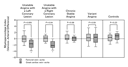
and Figure 2 <http://content.nejm.org/cgi/content/full/347/1/#F2> ). The
change in neutrophil myeloperoxidase content across the coronary circulation
was significantly greater than the difference in neutrophil myeloperoxidase
content between aortic and femoral venous blood in both patients with
unstable angina with a left coronary lesion and those with unstable angina
with a right coronary lesion, but not in any of the other three groups (
Table 1 <http://content.nejm.org/cgi/content/full/347/1/#T1> and Figure 2
<http://content.nejm.org/cgi/content/full/347/1/#F2> ).
<http://content.nejm.org/cgi/content/full/347/1/5/F2>
View larger version (18K):
[in this window] <http://content.nejm.org/cgi/content/full/347/1/5/F2>
[in a new window] <http://content.nejm.org/cgi/content-nw/full/347/1/5/F2>
Figure 2. VenousArterial Differences in Myeloperoxidase Content across the
Femoral and Coronary Vascular Beds.
Data are presented as medians, with 25th and 75 percentiles (boxes) and 10th
and 90th percentiles (I bars). The difference in myeloperoxidase content
across the coronary circulation was significantly greater in both patients
with unstable angina with a left coronary lesion and those with unstable
angina with a right coronary lesion than in patients with chronic stable
angina, patients with variant angina, and control patients. The difference
in myeloperoxidase content across the coronary vascular bed was
significantly greater than that across the femoral vascular bed in both
patients with unstable angina with a left coronary lesion and those with
unstable angina with a right coronary lesion, but not in any of the other
three groups. The asterisk indicates P<0.05 for the comparison of the group
with unstable angina with a left coronary lesion and unstable angina with a
right coronary lesion with the group with chronic stable angina, the group
with variant angina, and controls.
Correlation between Levels of C-Reactive Protein and Myeloperoxidase
The median plasma levels of C-reactive protein were similar in patients with
unstable angina with a left coronary lesion (6.5 mg per liter) and those
with unstable angina with a right coronary lesion (4.5 mg per liter, P=0.76)
and were significantly higher than the levels in patients with stable angina
(2.1 mg per liter), patients with variant angina (1.8 mg per liter), and
controls (1.2 mg per liter; P<0.01 for all comparisons) ( Table 1
<http://content.nejm.org/cgi/content/full/347/1/#T1> ).
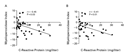
Overall, in the five groups, a significant correlation was found between
systemic levels of C-reactive protein and the aortic neutrophil
myeloperoxidase content (r=0.45, P=0.03), as well as between systemic
levels of C-reactive protein and the neutrophil myeloperoxidase content in
blood from the great cardiac vein (r=0.41, P=0.01) ( Figure 3
<http://content.nejm.org/cgi/content/full/347/1/#F3> ).
<http://content.nejm.org/cgi/content/full/347/1/5/F3>
View larger version (12K):
[in this window] <http://content.nejm.org/cgi/content/full/347/1/5/F3>
[in a new window] <http://content.nejm.org/cgi/content-nw/full/347/1/5/F3>
Figure 3. Correlation between Systemic Values for C-Reactive Protein and the
Change in the Myeloperoxidase Content in Blood from the Aorta and the Great
Cardiac Vein.
C-reactive protein values were significantly and inversely correlated with
the myeloperoxidase content of blood from the aorta (Panel A) and the great
cardiac vein (Panel B). The diagonal lines are the regression lines.
Discussion
Our findings confirm previous reports that in patients with unstable angina,
leukocytes become activated as they traverse the coronary vascular bed, 15
<http://content.nejm.org/cgi/content/full/347/1/#R15> , 16
<http://content.nejm.org/cgi/content/full/347/1/#R16> and that such
activation may be detectable systematically. 17
<http://content.nejm.org/cgi/content/full/347/1/#R17> , 23
<http://content.nejm.org/cgi/content/full/347/1/#R23> , 24
<http://content.nejm.org/cgi/content/full/347/1/#R24> In addition, we found
no significant increase in neutrophil activation in the great cardiac vein
in controls, in patients with stable angina and documented left anterior
descending coronary stenosis, or in patients with active variant angina and
recurrent ischemia in the territory of the left anterior descending coronary
artery. Moreover, there was no detectable increase in neutrophil activation
through the femoral circulation in any of the five groups studied.
In patients with unstable angina, transcoronary neutrophil activation was
not confined to the vascular bed perfused by the artery in which the culprit
stenosis was located and thus subjected to recurrent ischemia. In fact,
neutrophil activation occurred to a similar extent in patients in whom the
left anterior descending coronary artery was not the site of the culprit
stenosis. Patients with unstable angina and a culprit lesion in the right
coronary artery had only minimal atherosclerotic involvement of the left
anterior descending coronary artery, which was angiographically normal in
three patients, had only luminal irregularities in three patients, and had
stenosis of less than 50 percent of the diameter in three patients.
In animal models, neutrophil activation has been observed after 15 minutes
of coronary occlusionreperfusion. 25
<http://content.nejm.org/cgi/content/full/347/1/#R25> However, our findings
cannot be explained simply on the basis of an ischemiareperfusion
mechanism, in view of the fact that transcardiac neutrophil activation was
not observed in patients with active variant angina, spasm of the left
anterior descending artery, and a total ischemic burden similar to that of
patients with unstable angina.
In patients with unstable angina, inflammatory-cell infiltrates are commonly
found in most atherosclerotic plaques at postmortem examination 1
<http://content.nejm.org/cgi/content/full/347/1/#R1> and in endarterectomy
specimens. 2 <http://content.nejm.org/cgi/content/full/347/1/#R2> , 26
<http://content.nejm.org/cgi/content/full/347/1/#R26> Multiple fissured,
thrombosed coronary plaques seem to be a common finding in acute coronary
syndromes. Falk et al. reported 103 fissured, thrombosed plaques in 47
patients, 13 <http://content.nejm.org/cgi/content/full/347/1/#R13> and
Davies and Thomas reported 111 fissured, thrombosed plaques in 76 patients.
14 <http://content.nejm.org/cgi/content/full/347/1/#R14> Neither of these
reports discussed the possible clinical significance of the simultaneous
rupture of multiple plaques. Multiple plaques with inflammatory-cell
infiltrates and with a high content of proinflammatory cytokines were
reported by Arbustini et al. 12
<http://content.nejm.org/cgi/content/full/347/1/#R12> Finally, multiple
complex lesions were reported by Goldstein et al. 11
<http://content.nejm.org/cgi/content/full/347/1/#R11> The possibility that
multiple plaque fissures and thrombi develop simultaneously at different
sites merely as a result of mechanical stress seems rather unlikely. It
would appear more reasonable to speculate that a multifocal or widespread
inflammatory activation of the endothelium could change the characteristics
of the interface between the blood and the vessel walls from anticoagulant
and vasodilative to prothrombotic and vasoconstrictive, while at the same
time activating the metalloproteases and collagenases responsible for
endothelial-cell detachment and lysis of the plaque capsule at the sites
where it is weakest.
Whether neutrophils become activated by interacting with the surface of
sparse inflamed plaques or as a result of more widespread contact with a
diffusely inflamed coronary endothelium is not known. De Servi et al.
detected no activation of monocytes and neutrophils across the culprit
coronary stenosis in patients with unstable angina. 16
<http://content.nejm.org/cgi/content/full/347/1/#R16> Conversely, the
possibility of widespread coronary inflammation is suggested by the reports
of alterations in coronary flow 27
<http://content.nejm.org/cgi/content/full/347/1/#R27> , 28
<http://content.nejm.org/cgi/content/full/347/1/#R28> and [18F]deoxyglucose
uptake 29 <http://content.nejm.org/cgi/content/full/347/1/#R29> in
myocardial territories perfused by arteries without stenosis or culprit
lesions in patients with recent infarctions and in those with unstable
angina. Finally, in 10 percent of patients with unstable angina,
inflammatory red streaks were observed along nonstenosed coronary arteries
at the time of bypass surgery. 30
<http://content.nejm.org/cgi/content/full/347/1/#R30>
The reported prevalence of systemically detectable inflammatory markers in
acute coronary syndromes varies. Serum levels of C-reactive protein and of
proinflammatory cytokines such as interleukin-6 are elevated in about 70
percent of patients with severe unstable angina on admission, 31
<http://content.nejm.org/cgi/content/full/347/1/#R31> , 32
<http://content.nejm.org/cgi/content/full/347/1/#R32> in 50 percent of such
patients at discharge, and in 45 percent of such patients at six months of
follow-up. 20 <http://content.nejm.org/cgi/content/full/347/1/#R20> These
increased levels are associated with recurrent instability and acute
infarction. Accordingly, elevated levels of C-reactive protein and
interleukin-6 are found before the appearance of markers of myocardial
necrosis in nearly all patients in whom infarction is preceded by unstable
angina, but in less than 50 percent of patients with myocardial infarction
not preceded by unstable angina. 31
<http://content.nejm.org/cgi/content/full/347/1/#R31> , 33
<http://content.nejm.org/cgi/content/full/347/1/#R33> Therefore, the
triggers of coronary thrombosis and vasoconstriction are not necessarily the
same in all patients with acute coronary syndromes.
The activation of neutrophils as they traverse the coronary circulation of
patients with unstable angina is a marker of a widespread inflammatory
process occurring in the coronary vasculature. When the intensity of the
inflammatory stimuli varies, such a process may lead to waxing and waning of
thrombosis and vasoconstriction. The possibility of widespread coronary
inflammation has important implications for research and therapy. It
challenges the widely accepted hypothesis that a single vulnerable plaque is
responsible for the development of coronary instability a hypothesis that
is currently stimulating the development of techniques for the detection and
stabilization of such plaques.
Supported by grants from the National Research Council, Rome
(94.00518.PF41), the European Community (PL951505), and the Fondazione
Internazionale di Ricerca per il Cuore Onlus, Rome.
Source Information
From the Institute of Cardiology (A.B., L.M.B., G.L., F.C.) and the
Institute of Hematology (G.D.), Catholic University, Rome; and the
Cardiothoracic and Vascular Department, University Vita e Salute, Milan,
Italy (A.M.).
Address reprint requests to Dr. Maseri at the Cardiothoracic and Vascular
Department, University Vita e Salute, San Raffaele Hospital, Via Olgettina
60, 20132 Milan, Italy, or at [log in to unmask]
<mailto:[log in to unmask]> .
References
1. van der Wal AC, Becker AE, van der Loos CM, Das PK. Site of intimal
rupture or erosion of thrombosed coronary atherosclerotic plaques is
characterized by an inflammatory process irrespective of the dominant plaque
morphology. Circulation 1994;89:36-44. [Abstract]
<http://content.nejm.org/cgi/ijlink?linkType=ABST&journalCode=circulationaha
&resid=89/1/36>
2. Moreno PR, Falk E, Palacios IF, Newell JB, Fuster V, Fallon JT.
Macrophage infiltration in acute coronary syndromes: implications for plaque
rupture. Circulation 1994;90:775-778. [Abstract]
<http://content.nejm.org/cgi/ijlink?linkType=ABST&journalCode=circulationaha
&resid=90/2/775>
3. Libby P. Molecular bases of the acute coronary syndromes. Circulation
1995;91:2844-2850. [Full Text]
<http://content.nejm.org/cgi/ijlink?linkType=FULL&journalCode=circulationaha
&resid=91/11/2844>
4. Shah PK, Falk E, Badimon JJ, et al. Human monocyte-derived macrophages
induce collagen breakdown in fibrous caps of atherosclerotic plaques:
potential role of matrix-degrading metalloproteinases and implications for
plaque rupture. Circulation 1995;92:1565-1569. [ISI]
<http://content.nejm.org/cgi/external_ref?access_num=MULTIPLE_RESULTS&link_t
ype=ISI> [Medline]
<http://content.nejm.org/cgi/external_ref?access_num=7664441&link_type=MED>
5. Libby P. Current concepts of the pathogenesis of the acute coronary
syndromes. Circulation 2001;104:365-372. [Full Text]
<http://content.nejm.org/cgi/ijlink?linkType=FULL&journalCode=circulationaha
&resid=104/3/365>
6. Casscells W, Hathorn B, David M, et al. Thermal detection of cellular
infiltrates in living atherosclerotic plaques: possible implications for
plaque rupture and thrombosis. Lancet 1996;347:1447-1451. [ISI]
<http://content.nejm.org/cgi/external_ref?access_num=A1996UM48500011&link_ty
pe=ISI> [Medline]
<http://content.nejm.org/cgi/external_ref?access_num=8676628&link_type=MED>
7. Stefanadis C, Diamantopoulos L, Vlachopoulos C, et al. Thermal
heterogeneity within human atherosclerotic coronary arteries detected in
vivo: a new method of detection by application of a special thermography
catheter. Circulation 1999;99:1965-1971. [Abstract/Full Text]
<http://content.nejm.org/cgi/ijlink?linkType=ABST&journalCode=circulationaha
&resid=99/15/1965>
8. Fayad ZA, Fuster V, Fallon JT, et al. Noninvasive in vivo human coronary
artery lumen and wall imaging using black-blood magnetic resonance imaging.
Circulation 2000;102:506-510. [Abstract/Full Text]
<http://content.nejm.org/cgi/ijlink?linkType=ABST&journalCode=circulationaha
&resid=102/5/506>
9. Libby P. Coronary artery injury and the biology of atherosclerosis:
inflammation, thrombosis, and stabilization. Am J Cardiol 2000;86:Suppl
2:3-8. [Medline]
<http://content.nejm.org/cgi/external_ref?access_num=11074032&link_type=MED>
10. Rabbani R, Topol EJ. Strategies to achieve coronary arterial plaque
stabilization. Cardiovasc Res 1999;41:402-417. [ISI]
<http://content.nejm.org/cgi/external_ref?access_num=000079137800010&link_ty
pe=ISI> [Medline]
<http://content.nejm.org/cgi/external_ref?access_num=10341840&link_type=MED>
11. Goldstein JA, Demetriou D, Grines CL, Pica M, Shoukfeh M, O'Neill WW.
Multiple complex coronary plaques in patients with acute myocardial
infarction. N Engl J Med 2000;343:915-922. [Abstract/Full Text]
<http://content.nejm.org/cgi/ijlink?linkType=ABST&journalCode=nejm&resid=343
/13/915>
12. Arbustini E, Grasso M, Diegoli M, et al. Coronary atherosclerotic
plaques with and without thrombus in ischemic heart syndromes: a
morphologic, immunohistochemical, and biochemical study. Am J Cardiol
1991;68:Suppl:36B-50B. [Medline]
<http://content.nejm.org/cgi/external_ref?access_num=1892066&link_type=MED>
13. Falk E, Shah PK, Fuster V. Coronary plaque disruption. Circulation
1995;92:657-671. [Full Text]
<http://content.nejm.org/cgi/ijlink?linkType=FULL&journalCode=circulationaha
&resid=92/3/657>
14. Davies MJ, Thomas A. Thrombosis and acute coronary-artery lesions in
sudden cardiac ischemic death. N Engl J Med 1984;310:1137-1140. [Abstract]
<http://content.nejm.org/cgi/ijlink?linkType=ABST&journalCode=nejm&resid=310
/18/1137>
15. Mazzone A, De Servi S, Ricevuti G, et al. Increased expression of
neutrophil and monocyte adhesion molecules in unstable coronary artery
disease. Circulation 1993;88:358-363. [Abstract]
<http://content.nejm.org/cgi/ijlink?linkType=ABST&journalCode=circulationaha
&resid=88/2/358>
16. de Servi S, Mazzone A, Ricevuti G, et al. Expression of neutrophil and
monocyte CD11B/CD18 adhesion molecules at different sites of the coronary
tree in unstable angina pectoris. Am J Cardiol 1996;78:564-568. [ISI]
<http://content.nejm.org/cgi/external_ref?access_num=A1996VG43100015&link_ty
pe=ISI> [Medline]
<http://content.nejm.org/cgi/external_ref?access_num=8806345&link_type=MED>
17. Biasucci LM, D'Onofrio G, Liuzzo G, et al. Intracellular neutrophil
myeloperoxidase is reduced in unstable angina and myocardial infarction, but
its reduction is not related to ischemia. J Am Coll Cardiol 1996;27:611-616.
[ISI]
<http://content.nejm.org/cgi/external_ref?access_num=A1996TY10400013&link_ty
pe=ISI> [Medline]
<http://content.nejm.org/cgi/external_ref?access_num=8606272&link_type=MED>
18. Hazen SL, d'Avignon A, Anderson MM, Hsu FF, Heinecke JW. Human
neutrophils employ the myeloperoxidase-hydrogen peroxide-chloride system to
oxidize alpha-amino acids to a family of reactive aldehydes: mechanistic
studies identifying labile intermediates along the reaction pathway. J Biol
Chem 1998;273:4997-5005. [Abstract/Full Text]
<http://content.nejm.org/cgi/ijlink?linkType=ABST&journalCode=jbc&resid=273/
9/4997>
19. Ganz W, Tamura K, Marcus HS, Donoso R, Yoshida S, Swan HJ. Measurement
of coronary sinus blood flow by continuous thermodilution in man.
Circulation 1971;44:181-195. [ISI]
<http://content.nejm.org/cgi/external_ref?access_num=MULTIPLE_RESULTS&link_t
ype=ISI> [Medline]
<http://content.nejm.org/cgi/external_ref?access_num=4935053&link_type=MED>
20. Biasucci LM, Liuzzo G, Grillo RL, et al. Elevated levels of C-reactive
protein at discharge in patients with unstable angina predict recurrent
instability. Circulation 1999;99:855-860. [Abstract/Full Text]
<http://content.nejm.org/cgi/ijlink?linkType=ABST&journalCode=circulationaha
&resid=99/7/855>
21. Alternatives to analysis of variance and the t test based on ranks. In:
Glantz SA. Primer of biostatistics. 2nd ed. New York: McGraw-Hill,
1987:287-330.
22. Bogaty P, Brecker SJ, White SE, et al. Comparison of coronary
angiographic findings in acute and chronic first presentation of ischemic
heart disease. Circulation 1993;87:1938-1946. [Abstract]
<http://content.nejm.org/cgi/ijlink?linkType=ABST&journalCode=circulationaha
&resid=87/6/1938>
23. Mehta J, Dinerman J, Mehta P, et al. Neutrophil function in ischemic
heart disease. Circulation 1989;79:549-556. [Abstract]
<http://content.nejm.org/cgi/ijlink?linkType=ABST&journalCode=circulationaha
&resid=79/3/549>
24. Dinerman JL, Mehta JL, Saldeen TG, et al. Increased neutrophil elastase
release in unstable angina pectoris and acute myocardial infarction. J Am
Coll Cardiol 1990;15:1559-1563. [ISI]
<http://content.nejm.org/cgi/external_ref?access_num=A1990DH12900018&link_ty
pe=ISI> [Medline]
<http://content.nejm.org/cgi/external_ref?access_num=2345235&link_type=MED>
25. Jordan JE, Zhao ZQ, Vinten-Johansen J. The role of neutrophils in
myocardial ischemia-reperfusion injury. Cardiovasc Res 1999;43:860-878.
[ISI]
<http://content.nejm.org/cgi/external_ref?access_num=000082376700007&link_ty
pe=ISI> [Medline]
<http://content.nejm.org/cgi/external_ref?access_num=10615413&link_type=MED>
26. DiSciascio G, Cowley MJ, Goudreau E, Vetrovec GW, Johnson DE.
Histopathologic correlates of unstable ischemic syndromes in patients
undergoing directional coronary atherectomy: in vivo evidence of thrombosis,
ulceration, and inflammation. Am Heart J 1994;128:419-426. [ISI]
<http://content.nejm.org/cgi/external_ref?access_num=A1994PE86800001&link_ty
pe=ISI> [Medline]
<http://content.nejm.org/cgi/external_ref?access_num=8074000&link_type=MED>
27. Uren NG, Crake T, Lefroy DC, de Silva R, Davies GJ, Maseri A. Reduced
coronary vasodilator function in infarcted and normal myocardium after
myocardial infarction. N Engl J Med 1994;331:222-227. [Abstract/Full Text]
<http://content.nejm.org/cgi/ijlink?linkType=ABST&journalCode=nejm&resid=331
/4/222>
28. Gibson CM, Ryan KA, Murphy SA, et al. Impaired coronary blood flow in
nonculprit arteries in the setting of acute myocardial infarction. J Am Coll
Cardiol 1999;34:974-982. [ISI]
<http://content.nejm.org/cgi/external_ref?access_num=000082936900004&link_ty
pe=ISI> [Medline]
<http://content.nejm.org/cgi/external_ref?access_num=10520778&link_type=MED>
29. Araujo LI, Camici P, Spinks TJ, Jones T, Maseri A. Abnormalities in
myocardial metabolism in patients with unstable angina as assessed by
positron emission tomography. Cardiovasc Drugs Ther 1988;2:41-46. [Medline]
<http://content.nejm.org/cgi/external_ref?access_num=3154693&link_type=MED>
30. Wallsh E, Weinstein GS, Franzone A, Clavel A, Rossi PA, Kreps E.
Inflammation of the coronary arteries in patients with unstable angina. Tex
Heart Inst J 1986;13:105-108. [ISI]
<http://content.nejm.org/cgi/external_ref?access_num=A1986A983200015&link_ty
pe=ISI>
31. Liuzzo G, Biasucci LM, Gallimore JR, et al. The prognostic value of
C-reactive protein and serum amyloid A protein in severe unstable angina. N
Engl J Med 1994;331:417-424. [Abstract/Full Text]
<http://content.nejm.org/cgi/ijlink?linkType=ABST&journalCode=nejm&resid=331
/7/417>
32. Biasucci LM, Vitelli A, Liuzzo G, et al. Elevated levels of
interleukin-6 in unstable angina. Circulation 1996;94:874-877.
[Abstract/Full Text]
<http://content.nejm.org/cgi/ijlink?linkType=ABST&journalCode=circulationaha
&resid=94/5/874>
33. Liuzzo G, Biasucci LM, Gallimore JR, et al. Enhanced inflammatory
response in patients with preinfarction unstable angina. J Am Coll Cardiol
1999;34:1696-1703. [ISI]
<http://content.nejm.org/cgi/external_ref?access_num=000083778300009&link_ty
pe=ISI> [Medline]
<http://content.nejm.org/cgi/external_ref?access_num=10577559&link_type=MED>
Edward E. Rylander, M.D.
Diplomat American Board of Family Practice.
Diplomat American Board of Palliative Medicine.
|


